Introduction
Clinicians face significant challenges in interpreting T wave morphology, which can directly impact patient outcomes. Hyperacute T waves and peaked T waves are both significant indicators of underlying cardiac conditions, yet they present distinct characteristics that can influence treatment decisions. As healthcare professionals grapple with the challenge of accurately interpreting these patterns, the question arises: how can one effectively differentiate between these two T wave forms to ensure timely and appropriate interventions?
We will explore the key differences between hyperacute and peaked T waves, their clinical implications, and the advanced tools that can enhance diagnostic accuracy in urgent care settings across Canada.
Define Hyperacute T Waves and Peaked T Waves
Accurate ECG analysis is vital for identifying critical cardiac conditions, yet many challenges persist in recognizing early T signals. Hyperacute T waves vs peaked T waves are characterized by broad, asymmetrically peaked shapes, often appearing early in the progression of sudden myocardial infarction (MI). These patterns often show increased amplitude and can hide the ST segment, signaling serious cardiac distress. In Canada, traditional STEMI criteria often miss over 50% of acute coronary blockages, complicating timely diagnosis. Conversely, when comparing hyperacute T waves vs peaked T waves, the peaked T shapes are tall, narrow, and symmetrically peaked, commonly associated with hyperkalemia. These T patterns are frequently observed in individuals with elevated potassium levels and can indicate underlying electrolyte imbalances or cardiac issues. It's essential to grasp these definitions to effectively differentiate between hyperacute T waves vs peaked T waves in clinical practice. Moreover, distinguishing genuine very early T deflections from other irregularities is crucial for accurate diagnosis and treatment. The De-Winter T pattern, indicative of proximal left anterior descending (LAD) blockages, further underscores the importance of T patterns in diagnosing sudden coronary occlusions.
In this context, incorporating MaxYield™, an advanced automated ECG analysis platform from Neural Cloud Solutions Inc., can significantly improve the detection of early T signals. MaxYield™ maps ECG signals through noise, isolating and labeling key features in every heartbeat, which is vital for recognizing HATFs amidst noise and artifacts. As Dr. Alan Rabinowitz notes, "MaxYield™ rivals human interpretation in precision, ensuring that critical cardiac events are not overlooked." The platform's ability to deliver beat-by-beat analysis and its advanced noise filtering capabilities allow clinicians to salvage previously obscured sections of lengthy Holter and patch monitor recordings. This is especially crucial in urgent care environments where prompt and precise diagnosis can enhance individual outcomes. Using advanced tools like MaxYield™ can lead to better diagnostic accuracy, which is crucial for improving patient outcomes. Integrating insights from case studies can further demonstrate the diagnostic challenges and implications of hyperacute T patterns, ultimately enhancing individual outcomes in urgent care environments.

Compare Diagnostic Significance of Each T Wave Pattern
Detecting hyperacute T waves vs peaked T waves is crucial for timely intervention in acute coronary blockage, yet many healthcare professionals face challenges in identifying these vital signs. The distinction between hyperacute T waves vs peaked T waves serves as early indicators for myocardial infarction, prompting immediate interventions like reperfusion therapy, which are essential for improving patient outcomes. In contrast, the distinction between hyperacute T waves vs peaked T waves is primarily associated with hyperkalemia, indicating a need for electrolyte management rather than direct cardiac intervention. While both hyperacute T waves vs peaked T waves are clinically significant, elevated T forms demand more immediate attention due to their connection with acute coronary incidents.
Recent studies show that sharp T deflections can be identified in patients without conventional STEMI criteria. They have a specificity of 98.4% and a positive predictive value of 47.4%. This highlights their usefulness in emergency diagnostics. Moreover, the HATW score, which measures T-wave features, indicates that 49% of individuals diagnosed with obstructive myocardial ischemia had noticeable acute T waves three hours before angiographic confirmation. This underscores the importance of recognizing hyperacute T waves vs peaked T waves as a STEMI equivalent, prompting timely intervention to mitigate adverse cardiac events.
In urban areas of Canada, like Toronto and Calgary, using advanced algorithms to recognize T patterns can significantly improve diagnostic precision. This is crucial for enhancing care in urgent situations. Additionally, the integration of MaxYield™ by Neural Cloud Solutions Inc. enhances the efficiency of ECG analysis. MaxYield™ employs advanced noise filtering and unique signal recognition capabilities, enabling the identification and labeling of critical data even in recordings with high levels of noise and artifact. This is particularly beneficial in salvaging previously obscured sections of lengthy Holter, 1-Lead, and patch monitor recordings.
Experts such as Dr. Alan Rabinowitz and Dr. Brett Heilbron point out that MaxYield™ matches human interpretation in accuracy, leading to a significant boost in diagnostic yield. Consequently, applying MaxYield™ can enable faster and more precise evaluations of hyperacute T waves vs peaked T waves, ultimately leading to improved patient outcomes. Furthermore, with its FDA Class II SaMD clearance-pending status, MaxYield™ is positioned to meet regulatory standards, ensuring compliance and reliability in clinical settings.

Examine Morphological Characteristics of T Waves
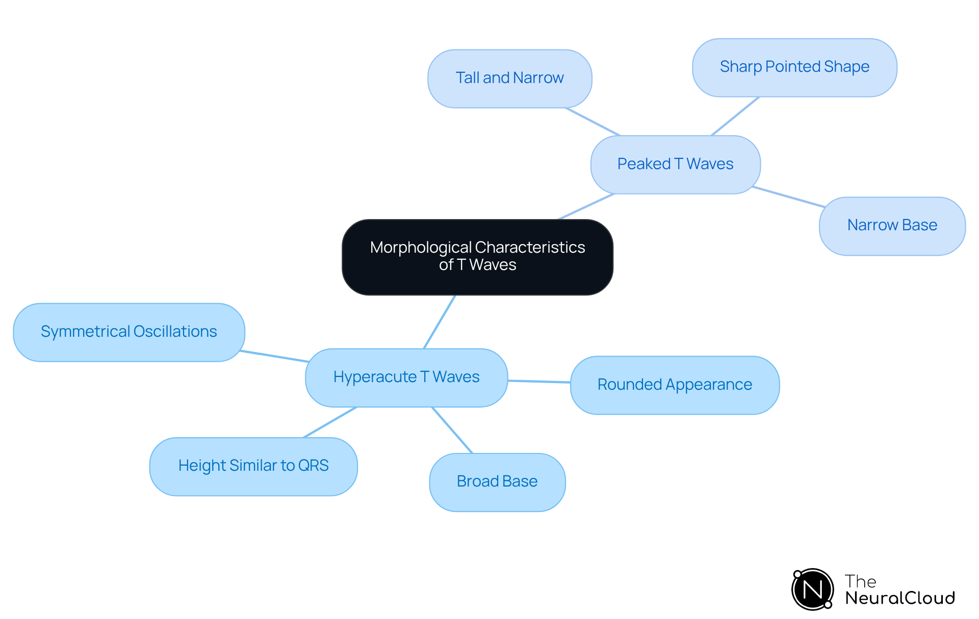
Accurate ECG analysis is critical, yet it often presents challenges that can lead to misdiagnosis. When comparing hyperacute T waves vs peaked T waves, hyperacute T forms have a broad base and rounded appearance, often reaching heights similar to or greater than the QRS complex. These oscillations are typically symmetrical and may obscure the ST segment, making them a vital indicator of acute ischemia. In contrast, when comparing hyperacute T waves vs peaked T waves, peaked T forms are tall, narrow, and sharply pointed, often described as tented. They arise from a narrow base and are also symmetrical, but their distinct shape sets them apart from sharp T forms. Understanding the differences between hyperacute T waves vs peaked T waves is essential for accurate diagnosis and effective management in clinical practice.
Recent Canadian research emphasizes the importance of identifying sharp T deflections as the earliest electrocardiographic indication of acute ischemia, occurring after QT prolongation and before ST-segment elevation. This distinction is crucial, especially in emergency settings where timely intervention can significantly impact patient outcomes. Case studies have shown that sharp T patterns can be misinterpreted if not thoroughly examined, particularly when distinguishing them from conditions like hyperkalemia, which can also alter T patterns. Therefore, clinicians must be skilled at recognizing these subtle differences to avoid misdiagnosis and ensure appropriate treatment.
To improve the precision of ECG analysis, health tech developers can utilize Neural Cloud Solutions Inc.'s MaxYield platform, which automates the detection of essential characteristics in ECG signals, including hyperacute T patterns. With MaxYield's AI-driven analysis, noise is minimized, and workflow efficiency is enhanced, allowing for rapid beat-by-beat assessments of up to 200,000 heartbeats in less than 5 minutes. As Dr. Alan Rabinowitz states, "MaxYield's precision rivals human interpretation, ensuring that subtle changes in T shape morphology are accurately captured." This capability is vital for clinicians in emergency settings, where every second counts. By integrating MaxYield into their platforms, healthcare professionals can transform noisy recordings into clear, actionable insights, ultimately leading to better patient outcomes. By mastering these distinctions, clinicians can significantly enhance patient care and outcomes.
Analyze Clinical Implications and Management Strategies
The presence of hyperacute T waves vs peaked T waves in ECG readings signals an urgent need for clinical intervention, often leading to treatments like thrombolysis or percutaneous coronary intervention to restore blood flow and prevent myocardial damage. When comparing hyperacute T waves vs peaked T waves, it is important to note that peaked T waves require a different approach that targets the underlying hyperkalemia, often necessitating treatments like calcium gluconate, insulin, or diuretics. Understanding these clinical implications allows healthcare providers to tailor their management strategies effectively, leading to better health outcomes for patients.
Furthermore, integrating wearable technology with Neural Cloud Solutions Inc.'s MaxYield™ platform enhances ECG analysis efficiency by automating labeling and data extraction. This integration not only streamlines processes but also enhances diagnostic accuracy, ultimately improving patient care. As Dr. Alan Rabinowitz notes, "MaxYield’s precision rivals human interpretation, significantly reducing noise and improving diagnostic confidence."
For clinicians in Toronto and Calgary, this capability is vital, as quick and accurate ECG analysis can truly impact patient care.

Conclusion
Differentiating between hyperacute and peaked T waves is crucial for effective ECG interpretation and patient management. These two T wave patterns, while both indicative of underlying cardiac issues, signal different conditions that require distinct management strategies. Misinterpreting these patterns can lead to inappropriate management and worsen patient outcomes. Recognizing these differences can significantly enhance diagnostic precision and improve patient outcomes in urgent care settings.
The article highlights the critical characteristics of hyperacute T waves, which often indicate acute myocardial infarction, and peaked T waves, commonly associated with hyperkalemia. It emphasizes the importance of advanced tools like MaxYield™ from Neural Cloud Solutions Inc., which can aid clinicians in accurately identifying these T wave patterns amidst noise and artifacts. Using this technology helps healthcare professionals catch vital signs that might otherwise be missed, leading to better management of cardiac conditions.
In conclusion, differentiating between hyperacute and peaked T waves is crucial, with significant implications for patient care. Clinicians in Canada, particularly in urban centers like Toronto and Calgary, are encouraged to adopt advanced ECG analysis tools to enhance their diagnostic capabilities. By doing so, they can improve the accuracy of their assessments and ensure timely interventions, thereby safeguarding patient health in critical situations. Embracing advanced ECG analysis tools can transform patient care and outcomes in critical situations.
Frequently Asked Questions
What are hyperacute T waves and peaked T waves?
Hyperacute T waves are characterized by broad, asymmetrically peaked shapes that often appear early in the progression of sudden myocardial infarction (MI). Peaked T waves, on the other hand, are tall, narrow, and symmetrically peaked, commonly associated with hyperkalemia.
Why is accurate ECG analysis important?
Accurate ECG analysis is vital for identifying critical cardiac conditions, as many challenges persist in recognizing early T signals. Misinterpretation can lead to missed diagnoses, particularly in acute coronary blockages.
How do hyperacute T waves and peaked T waves differ in their clinical significance?
Hyperacute T waves signal serious cardiac distress and can hide the ST segment, while peaked T waves indicate elevated potassium levels and may suggest underlying electrolyte imbalances or cardiac issues.
What is the impact of traditional STEMI criteria in Canada?
Traditional STEMI criteria often miss over 50% of acute coronary blockages in Canada, complicating timely diagnosis and treatment of cardiac conditions.
What is the De-Winter T pattern?
The De-Winter T pattern is indicative of proximal left anterior descending (LAD) blockages and highlights the importance of T patterns in diagnosing sudden coronary occlusions.
How can MaxYield™ improve ECG analysis?
MaxYield™, an advanced automated ECG analysis platform from Neural Cloud Solutions Inc., enhances the detection of early T signals by mapping ECG signals through noise, isolating and labeling key features in every heartbeat.
What advantages does MaxYield™ offer in urgent care environments?
MaxYield™ provides beat-by-beat analysis and advanced noise filtering capabilities, allowing clinicians to salvage previously obscured sections of lengthy Holter and patch monitor recordings, which is crucial for prompt and precise diagnosis.
How does improved diagnostic accuracy affect patient outcomes?
Using advanced tools like MaxYield™ can lead to better diagnostic accuracy, which is essential for improving patient outcomes in urgent care settings.
List of Sources
- Define Hyperacute T Waves and Peaked T Waves
- ECG Diagnosis: Hyperacute T Waves - PMC (https://pmc.ncbi.nlm.nih.gov/articles/PMC4500486)
- STEMI | Department of Emergency Medicine | Saint John (https://emsj.ca/tag/stemi)
- theneuralcloud.com (https://theneuralcloud.com/post/what-causes-peaked-t-waves-understanding-their-clinical-significance)
- Hyperacute T-Waves: ECG Signs of Early MI (https://powerfulmedical.com/blog/hyperacute-t-waves)
- Compare Diagnostic Significance of Each T Wave Pattern
- Hyperacute T-Waves: ECG Signs of Early MI (https://powerfulmedical.com/blog/hyperacute-t-waves)
- Hyperacute T Waves Are Specific for Occlusion Myocardial Infarction, Even Without Diagnostic ST-Segment Elevation - PMC (https://pmc.ncbi.nlm.nih.gov/articles/PMC12791876)
- Hyperacute T waves and Occlusion MI | ECG Cases| Emergency Medicine Cases (https://emergencymedicinecases.com/hyperacute-t-waves-occlusion-mi)
- ECG Diagnosis: Hyperacute T Waves | The Permanente Journal (https://thepermanentejournal.org/doi/10.7812/TPP/14-243)
- Actually, Hyperacute T Waves Can Help Identify Occlusion MI - JournalFeed (https://journalfeed.org/article-a-day/2023/actually-hyperacute-t-waves-can-help-identify-occlusion-mi)
- Examine Morphological Characteristics of T Waves
- ECG Diagnosis: Hyperacute T Waves - PMC (https://pmc.ncbi.nlm.nih.gov/articles/PMC4500486)
- Impending Danger: Evolution of Hyperacute T Waves to Cardiac Arrest (https://cureus.com/articles/432103-impending-danger-evolution-of-hyperacute-t-waves-to-cardiac-arrest)
- annemergmed.com (https://annemergmed.com/article/S0196-0644(22)01327-0/fulltext)
- Actually, Hyperacute T Waves Can Help Identify Occlusion MI - JournalFeed (https://journalfeed.org/article-a-day/2023/actually-hyperacute-t-waves-can-help-identify-occlusion-mi)
- Analyze Clinical Implications and Management Strategies
- Hyperacute T Waves Are Specific for Occlusion Myocardial Infarction, Even Without Diagnostic ST-Segment Elevation - PMC (https://pmc.ncbi.nlm.nih.gov/articles/PMC12791876)
- Hyperkalemia-Peaked T waves and ECG Recognition - Medcram Blog (https://blog.medcram.com/cme-courses/hyperkalemia-peaked-t-waves-and-ecg-recognition)
- Hyperkalemia Treatment & Management: Approach Considerations, Initial Emergency Management, Pharmacologic Therapy and Dialysis (https://emedicine.medscape.com/article/240903-treatment)
- Hyperkalemia – Diagnosis and Treatment : Emergency Care BC (https://emergencycarebc.ca/clinical_resource/clinical-summary/hyperkalemia-diagnosis-and-treatment)