Introduction
Navigating the complexities of ECG layout is crucial for health tech developers striving to enhance patient care through precise cardiac assessments. Let’s explore how to execute a flawless ECG layout step by step, focusing on best practices that can boost diagnostic precision.
Navigating the complexities of ECG layout can be daunting due to frequent issues like electrode misplacement and signal interference. To ensure accurate cardiac assessments, developers must adopt best practices that mitigate these common pitfalls.
Understand the Basics of ECG Layout
To effectively analyze ECGs, it's crucial to grasp the fundamental components of the EKG layout. An ECG assesses the heart's electrical activity via sensors positioned on the skin. The standard 12-channel EKG layout includes 10 sensors: four limb connections (right arm, left arm, right leg, left leg) and six precordial connections (V1 to V6) placed on the chest. Each electrode provides a unique viewpoint of the heart's electrical activity, enabling a comprehensive analysis.
- Learn the Waveforms: Familiarize yourself with the P wave, QRS complex, and T wave. The P wave shows atrial depolarization. The QRS complex indicates ventricular depolarization. Finally, the T wave represents ventricular repolarization.
- Recognize Lead Placement: Understand the anatomical landmarks for electrode placement. For example, V1 is positioned in the fourth intercostal space at the right sternal border, while V2 is placed at the same level on the left side. The correct placement of the EKG layout is crucial. Improper positioning within the EKG layout can lead to inaccuracies, complicating patient treatment.
- Understand Electrical Interpretation: Each lead captures unique electrical impulses that can indicate various cardiac conditions. Understanding these indicators is key to accurate diagnosis and effective treatment planning, particularly when considering the EKG layout. Recent advancements in ECG technology have improved the accuracy of signal interpretation, allowing for better patient outcomes.
- Review Common Artifacts: Be aware of potential artifacts that can distort ECG readings, such as muscle tremors or electrical interference. Being aware of these artifacts helps troubleshoot and ensures accurate readings. Research highlights that improper placement of the sensors can introduce artifacts, emphasizing the need for proper training and adherence to best practices in positioning.
By mastering these elements, healthcare professionals can significantly improve patient care through precise ECG analysis.

Prepare the Patient and Equipment for ECG Placement
Preparing for an ECG test is crucial, as improper preparation can lead to inaccurate results and patient discomfort. To ensure a successful ECG test, follow these preparation steps:
- Create a Comfortable Environment: Set the room temperature to a comfortable level and eliminate distractions. A calm environment helps the patient relax, which is crucial for minimizing movement artifacts during the test.
- Patient Positioning: Make sure the patient is lying flat on their back with their arms relaxed at their sides. This optimal positioning facilitates precise placement of sensors and reduces muscle tension, enhancing the quality of the ECG readings.
- Skin Preparation: Thoroughly clean the skin where sensors will be placed using an alcohol wipe to remove oils and dirt, ensuring optimal contact. If the patient has excessive body hair, shaving the area may be necessary to improve adhesion and signal quality.
- Check Equipment: Verify that the ECG machine is functioning correctly. Examine all connectors for soundness and ensure wire strands are in good condition. Familiarize yourself with the machine's settings to ensure proper configuration for the test.
- Explain the Procedure: Clearly communicate with the patient about what to expect during the ECG. Explain the test's purpose and reassure them that it is painless, which can significantly alleviate any anxiety they may have.
Following these best practices not only boosts patient comfort but also ensures reliable ECG results, which is vital for quality patient care.

Execute Proper Electrode Placement for 12-Lead ECG
Accurate ECG readings hinge on proper lead placement, a task that can be challenging even for experienced professionals. Follow these steps to execute correct placement:
-
Identify Lead Locations: Familiarize yourself with the locations of the 10 electrodes:
- Limb Leads: Place RA (right arm) and LA (left arm) leads on the deltoids, and RL (right leg) and LL (left leg) leads on the lower abdomen near the iliac crests.
- Precordial Leads: Place V1 in the fourth intercostal space at the right sternal border, V2 at the same level on the left side, V3 between V2 and V4, V4 in the fifth intercostal space at the midclavicular line, V5 at the anterior axillary line at the same level as V4, and V6 at the midaxillary line at the same level as V4.
-
Use Symmetrical Placement: Ensure that limb electrodes are positioned symmetrically to avoid misinterpretation. For example, the right arm electrode should be positioned on the right side and the left arm electrode on the left side. Did you know that nurses misplace ECG electrodes 64% of the time? This high rate of misplacement underscores the critical need for proper training and adherence to established protocols.
-
Check for Proper Adhesion: After placing the electrodes, ensure they are securely attached to the skin. Weak adhesion can result in artifacts in the readings, which can be reduced by the advanced noise filtering features of MaxYield™, ensuring clearer outputs.
-
Verify Connections: Double-check that all wire connections are secured properly to the ECG machine. Loose connections can result in flat lines or poor signal quality.
-
Instruct the Patient: Ask the patient to remain still and relaxed during the ECG recording. Movement can introduce artifacts that compromise the quality of the data. It's essential to recognize that cardiologists accurately position leads V1 and V2 in fewer than 20% of instances. This statistic highlights the urgent need for improved techniques and tools in ECG lead placement.
By integrating these steps, healthcare professionals can significantly improve the precision of ECG readings. This, in turn, enhances patient outcomes, particularly when combined with the features of MaxYield™, which additionally provides automated ECG labeling and data extraction to simplify the process.

Troubleshoot Common Issues in ECG Layout
ECG analysis can be challenging due to various technical issues that compromise the accuracy of readings. Here are common problems and their solutions:
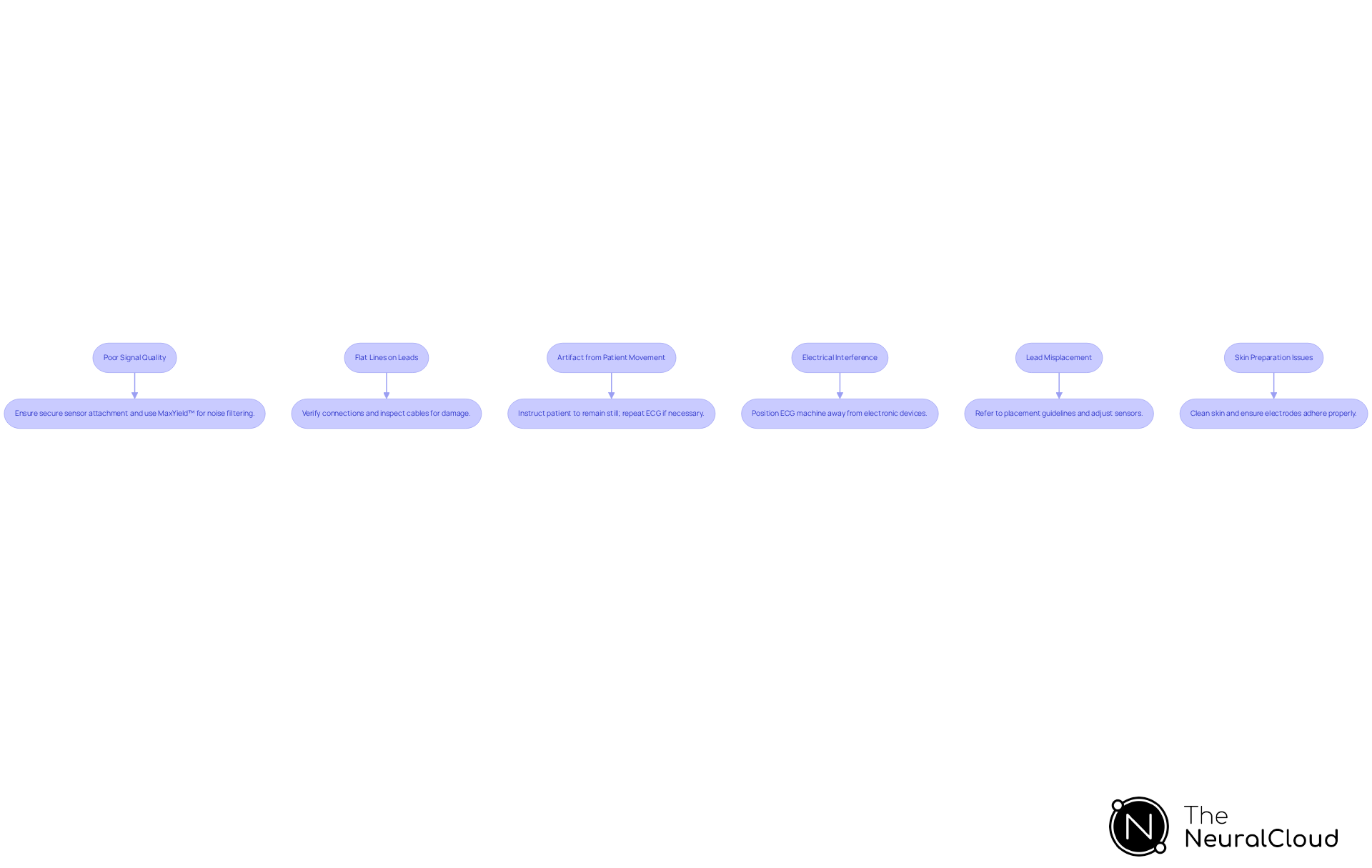
- Poor Signal Quality: Noisy or unclear ECG tracings often arise from improper placement of the sensors. Research shows that inadequate connector attachment can result in considerable distortion, affecting diagnostic precision. Ensure the devices are securely attached. Consider re-cleaning the skin with an alcohol wipe before reapplying them. Utilizing Neural Cloud Solutions' MaxYield™ platform can enhance signal quality through advanced noise filtering, ensuring that only clean signals are processed.
- Flat Lines on Leads: Flat lines may suggest disconnection of lead wires or inadequate contact with the sensors. Verify that all connections are secure and that electrodes are properly adhered to the skin. Regular inspection of cables for visible damage can prevent these issues.
- Artifact from Patient Movement: Patient movement during the test can introduce artifacts. Instruct the patient to remain still and relaxed. If movement occurs, it may be necessary to repeat the ECG to ensure accurate results. MaxYield™ can automate the evaluation of quality, assisting in recognizing and reducing the effect of such artifacts.
- Electrical Interference: External electrical sources can interfere with ECG readings. Position the ECG machine away from other electronic devices and ensure the environment is free from electromagnetic interference, which can compromise signal quality. Notably, AC interference can produce a fuzzy baseline on the ECG trace, so monitoring for this is essential.
- Lead Misplacement: Abnormal ECG results may be due to lead misplacement. Refer to standard placement guidelines to ensure accuracy. If necessary, make adjustments to the sensors and repeat the ECG to ensure accurate results.
- Skin Preparation Issues: If sensors are not adhering properly, ensure the skin is clean, dry, and free from oils. Proper skin preparation is essential; use an alcohol wipe to ready the skin before electrode placement to improve adhesion and quality.
By addressing these common issues, healthcare professionals can enhance the reliability of ECG readings. This improvement ultimately leads to better patient outcomes in clinical settings. Incorporating automated signal quality assessment methods, such as those offered by MaxYield™, can further ensure that only noise-free signals are used in decision-making systems, aligning with current trends in healthcare technology. Additionally, it is important to note that MaxYield™ is currently pending FDA Class II SaMD clearance, ensuring compliance with regulatory standards. Expert endorsements from Dr. Alan Rabinowitz, Dr. Brett Heilbron, and Dr. Marc W. Deyell emphasize MaxYield’s precision rivaling human interpretation and its noise reduction benefits. Furthermore, Insight360 serves as a complementary tool that transforms MaxYield’s clean ECG data into interactive dashboards and clinical-ready reports.
Conclusion
Accurate ECG analysis is often hindered by challenges such as improper electrode placement and patient discomfort. By understanding the intricacies of electrode placement, patient preparation, and troubleshooting common issues, professionals can significantly improve the reliability of ECG readings. This knowledge aids in precise diagnosis and fosters better treatment outcomes for patients.
Key insights from this guide emphasize:
- The importance of proper electrode placement
- The need for a comfortable patient environment
- The critical role of equipment checks
Recognizing common pitfalls, such as lead misplacement and electrical interference, allows healthcare providers to implement effective solutions that enhance the quality of ECG data. Utilizing advanced tools like MaxYield™ can further streamline the process, ensuring that healthcare professionals have access to the highest quality signals for interpretation.
In conclusion, mastering EKG layout is not just a technical skill; it is a vital component of delivering exceptional healthcare. By adhering to best practices and leveraging innovative technologies, healthcare professionals can ensure that every ECG reading contributes to better patient care and outcomes.
Frequently Asked Questions
What is the purpose of an ECG?
An ECG assesses the heart's electrical activity through sensors positioned on the skin, providing insights into cardiac function.
What does the standard 12-channel EKG layout consist of?
The standard 12-channel EKG layout includes 10 sensors: four limb connections (right arm, left arm, right leg, left leg) and six precordial connections (V1 to V6) placed on the chest.
What are the key waveforms to understand in an ECG?
The key waveforms include the P wave, which shows atrial depolarization; the QRS complex, which indicates ventricular depolarization; and the T wave, which represents ventricular repolarization.
How important is lead placement in ECG analysis?
Lead placement is crucial, as improper positioning can lead to inaccuracies in the ECG readings, complicating patient treatment.
Where are the V1 and V2 electrodes placed?
V1 is positioned in the fourth intercostal space at the right sternal border, while V2 is placed at the same level on the left side.
What is the significance of understanding electrical interpretation in ECGs?
Each lead captures unique electrical impulses that can indicate various cardiac conditions, making it essential for accurate diagnosis and effective treatment planning.
What are common artifacts that can affect ECG readings?
Common artifacts include muscle tremors and electrical interference, which can distort ECG readings and lead to inaccuracies.
Why is proper training important in ECG sensor placement?
Proper training is essential to adhere to best practices in positioning, as improper placement can introduce artifacts and compromise the accuracy of ECG readings.
List of Sources
- Understand the Basics of ECG Layout
- The latest innovations in ECG technology and analysis - Norav Medical (https://noravmedical.com/advancements-in-ecg-technology-exploring-the-latest-innovations-in-ecg-analysis)
- Accuracy of ECG chest electrode placements by paramedics: an observational study - PMC (https://pmc.ncbi.nlm.nih.gov/articles/PMC8312365)
- ECG signal feature extraction trends in methods and applications - BioMedical Engineering OnLine (https://link.springer.com/article/10.1186/s12938-023-01075-1)
- Prepare the Patient and Equipment for ECG Placement
- 12-Lead ECG Placement: The Ultimate Guide | Cables and Sensors (https://cablesandsensors.com/pages/12-lead-ecg-placement-guide-with-illustrations?srsltid=AU7gw4UpXnWVdmjFimL7z0au91pnMErkTBomXJQmJ2dbxRe5ZisfdsGL)
- Best Practices for ECG Lead Placement and Patient Preparation (https://nasiff.com/best-practices-for-ecg-lead-placement-and-patient-preparation)
- How to Prepare a Patient for an ECG Test | Midmark Diagnostic Training (https://midmark.com/service-support/technical-support/online-training/diagnostic-knowledge-base/how-to---prepare-a-patient-for-an-ecg-test)
- Execute Proper Electrode Placement for 12-Lead ECG
- Were your 12-lead ECG electrodes placed where they should be? (https://myheartsisters.org/2023/01/29/misplaced-ecg-electrodes)
- 12-Lead ECGs: Room for Improvement (https://jems.com/patient-care/12-lead-ecgs-room-for-improvement)
- Location, location, location: Electrode placement matters (https://ems1.com/medical-monitoring/location-location-location-electrode-placement-matters)
- Accuracy of ECG chest electrode placements by paramedics: an observational study - PMC (https://pmc.ncbi.nlm.nih.gov/articles/PMC8312365)
- What You Need to Know About ECG Electrode Placement in 2025 - INTCO Medical (https://intcomedical.com/news/what-you-need-to-know-about-ecg-electrode-placement-in-2025)
- Troubleshoot Common Issues in ECG Layout
- How To Troubleshoot Common ECG Machine Failures (https://allstatesmed.com/blogs/news/how-to-troubleshoot-common-ecg-machine-failures?srsltid=AU7gw4X5vvgzIQj-doFAGiVjWxTedzuKkeVxY2_cEgsx-4_ANc2H1vXS)
- ECG Signal Quality: A Practical Guide for ECG Readings (https://gehealthcare.com/en-gb/insights/article/ecg-signal-quality-a-practical-guide-for-ecg-readings?srsltid=AU7gw4XRngBVHJ0O-jmWmtkUMTfUkn9W2AyxC4FseXRO_MS-pOZgoSbw)
- Robustness of electrocardiogram signal quality indices - PMC (https://pmc.ncbi.nlm.nih.gov/articles/PMC9006023)
- ECG Leads Off Alarm: How to Fix It in 6 Steps (https://med-linket-corp.com/blogs/news/ecg-leads-off-alarm-fix?srsltid=AU7gw4VoibdSYiwNbseRt-wEZeOFKOhPmVRbi158yfJYcJaxNRxgiY3G)
- Reducing ECG artifact (https://numed.co.uk/news/reducing-ecg-artifact)