Introduction
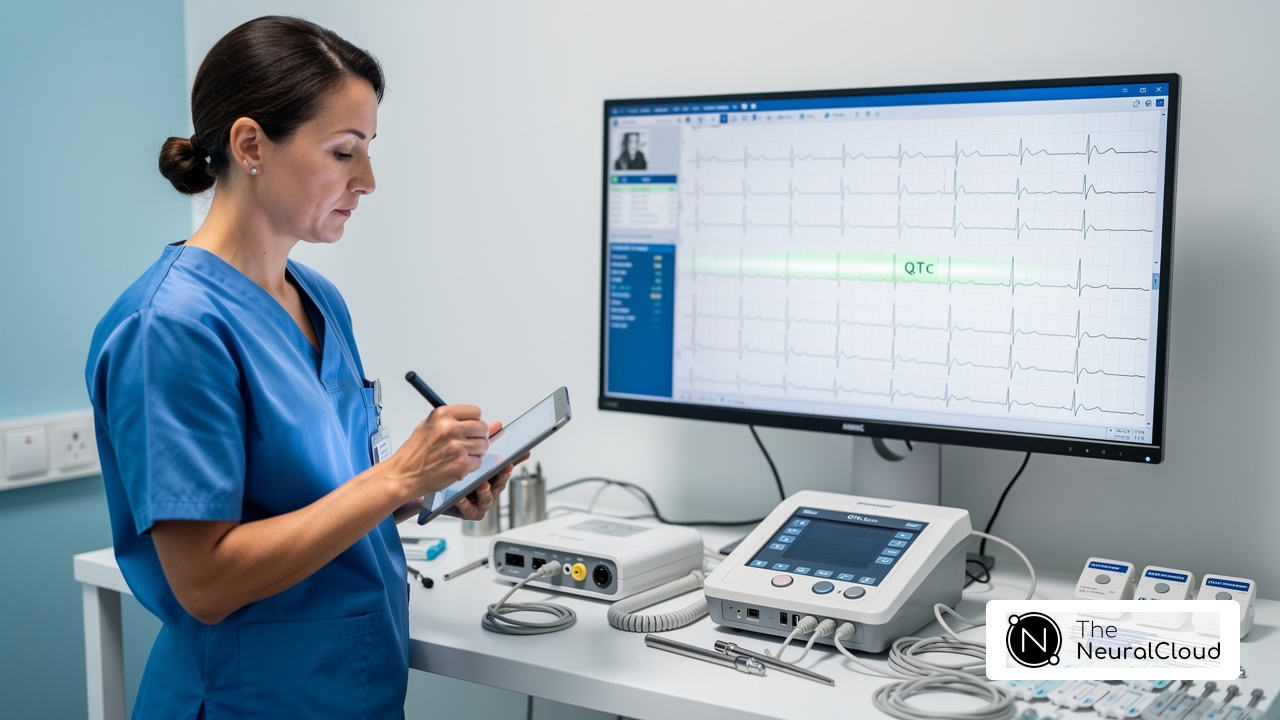
Healthcare professionals face significant challenges in interpreting normal sinus rhythm (NSR) with ST elevation in ECGs. This guide will help clinicians and researchers master how to identify and analyze NSR, leading to accurate diagnoses and timely interventions. Misinterpretations can lead to critical errors in patient care, making it essential to differentiate between benign ST elevation and serious cardiac events. Understanding these nuances is essential for effective patient management.
Define Normal Sinus Rhythm and Its Characteristics
Interpreting ECG data accurately is essential, yet many healthcare professionals face challenges due to noise and inaccuracies in recordings. Normal Sinus Rhythm (NSR) is characterized by electrical impulses originating from the sinoatrial (SA) node, resulting in a consistent heart rhythm. The key characteristics of NSR include:
- Heart Rate: Typically ranges from 60 to 100 beats per minute (bpm), which is considered normal for adults.
- P Waves: Each P wave precedes a QRS complex, indicating effective atrial depolarization. The P waves must be round, identical in shape, and present before every QRS complex in a 1:1 ratio.
- PR Interval: The interval between the P wave and the QRS complex is consistent, usually between 0.12 and 0.20 seconds, reflecting normal conduction through the atrioventricular node.
- QRS Complex: The QRS complex is narrow and uniform, indicating normal ventricular depolarization, typically less than 0.12 seconds in duration.
- Rhythm Regularity: The R-R intervals are consistent, demonstrating a stable rhythm with less than 0.16-second variation between successive P waves.
Grasping these characteristics helps healthcare professionals distinguish normal sinus rhythm with ST elevation from other cardiac rhythms, particularly in the context of ST elevation analysis. In Canadian patients, adherence to these criteria is essential for accurate diagnosis and treatment, as variations in heart rhythm can significantly impact clinical outcomes. Case studies have shown that recognizing the nuances of NSR can lead to improved management of cardiovascular conditions, reinforcing the importance of precise ECG interpretation in clinical practice.
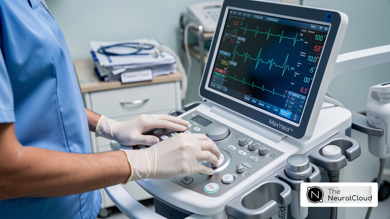
The MaxYield™ platform from Neural Cloud Solutions Inc. makes interpreting NSR easier by addressing the garbage-in/garbage-out issue associated with PPG feeds. By providing hospital-grade ECG accuracy from consumer-grade devices, MaxYield™ ensures clean data across all connected devices. Its advanced AI-driven analysis transforms noisy recordings into detailed insights, delivering beat-by-beat analysis of 200,000 heartbeats in less than 5 minutes. This capability not only reduces error variance in downstream models but also supports confident clinical decisions.
Expert endorsements from Dr. Alan Rabinowitz, Dr. Brett Heilbron, and Dr. Marc W. Deyell emphasize MaxYield’s precision, rivaling human interpretation and significantly reducing noise in ECG data. Furthermore, with FDA Class II SaMD clearance-pending status and audit-ready documentation supporting GxP inspections, MaxYield™ stands as a reliable tool for clinicians and researchers alike. Specific case studies have demonstrated that recognizing the nuances of NSR through MaxYield™ can lead to improved management of cardiovascular conditions, reinforcing the importance of precise ECG interpretation in clinical practice. With MaxYield™, clinicians can trust their ECG interpretations, ultimately leading to better patient outcomes and more effective cardiovascular management.

Explain ST Elevation: Clinical Significance and Interpretation
ST Elevation on an ECG presents significant challenges in accurate diagnosis and timely intervention for myocardial injury or ischemia. Its clinical significance includes:
- Indication of Myocardial Infarction: ST elevation is a hallmark of ST-Elevation Myocardial Infarction (STEMI), indicating full-thickness myocardial damage due to coronary artery occlusion. In Canada, recognizing ST rise in myocardial infarction cases is crucial for timely intervention, as quick identification can greatly enhance outcomes for individuals.
- Measurement Criteria: ST rise is considered significant if it surpasses 1 mm in two adjacent limb leads or 2 mm in chest leads V2-V3. These thresholds are essential for accurate diagnosis and treatment planning, guiding clinicians in identifying patients who require urgent care.
- Differential Diagnosis: While ST rise typically indicates ischemia, it can also arise from conditions such as pericarditis or early repolarization. Grasping these nuances is vital for accurate diagnosis. Misinterpretation can result in unnecessary interventions or delays in the right treatment.
Getting ST changes right is crucial for improving patient outcomes, making it a vital skill for healthcare professionals. Recent studies emphasize the importance of recognizing atypical presentations of myocardial infarction, which can sometimes mimic classic STEMI patterns. This underscores the need for thorough clinical evaluation and ECG interpretation.

Guide Through ECG Analysis: Steps to Identify Normal Sinus Rhythm with ST Elevation
Accurate identification of normal sinus rhythm with ST elevation on an ECG is crucial for effective patient management. To achieve this, follow these steps:
- Obtain a 12-channel ECG: Make sure the electrodes are placed correctly to get a complete picture of the heart's electrical activity. Studies show that accurate electrode placement is vital, as only 5.8% of participants in a recent observational study managed to place all chest electrodes correctly.
- Assess Heart Rate: Calculate the heart rate by counting the number of R-R intervals in a 6-second strip and multiplying by 10. Check that the heart rate is between 60 and 100 bpm to confirm normal sinus rhythm with ST elevation, as this range defines NSR.
- Evaluate P Waves: Look for P waves before each QRS complex. They should be uniform in shape and consistent in timing, indicating normal atrial depolarization.
- Measure PR Interval: Ensure the PR interval is between 0.12 and 0.20 seconds, which indicates normal conduction from the atria to the ventricles. A prolonged PR interval may suggest atrioventricular block.
- Analyze QRS Complex: Confirm that the QRS complexes are narrow and consistent, indicating normal ventricular depolarization. A duration of 0.06 to 0.10 seconds is considered normal.
- Identify ST Segment: Look for ST segment rise, especially in leads associated with the area of the heart being evaluated. Measure the height from the baseline at the J point, noting that ST heights greater than 1 mm in limb leads or 2 mm in chest leads are significant.
- Correlate with Clinical Context: Consider the patient's symptoms and history to determine if the ST rise indicates myocardial infarction or another condition. In cases of coronary vasospasm, ST changes may resolve with symptom resolution, highlighting the importance of clinical correlation.
Following this systematic approach helps ensure a thorough ECG analysis, leading to accurate diagnoses and timely interventions, which is essential in Canadian healthcare settings where precise cardiac assessments are critical.

Identify Common Misinterpretations in ST Elevation Analysis
Misinterpretations in ST rise analysis can lead to significant diagnostic challenges, causing delays in appropriate treatment. Key pitfalls include:
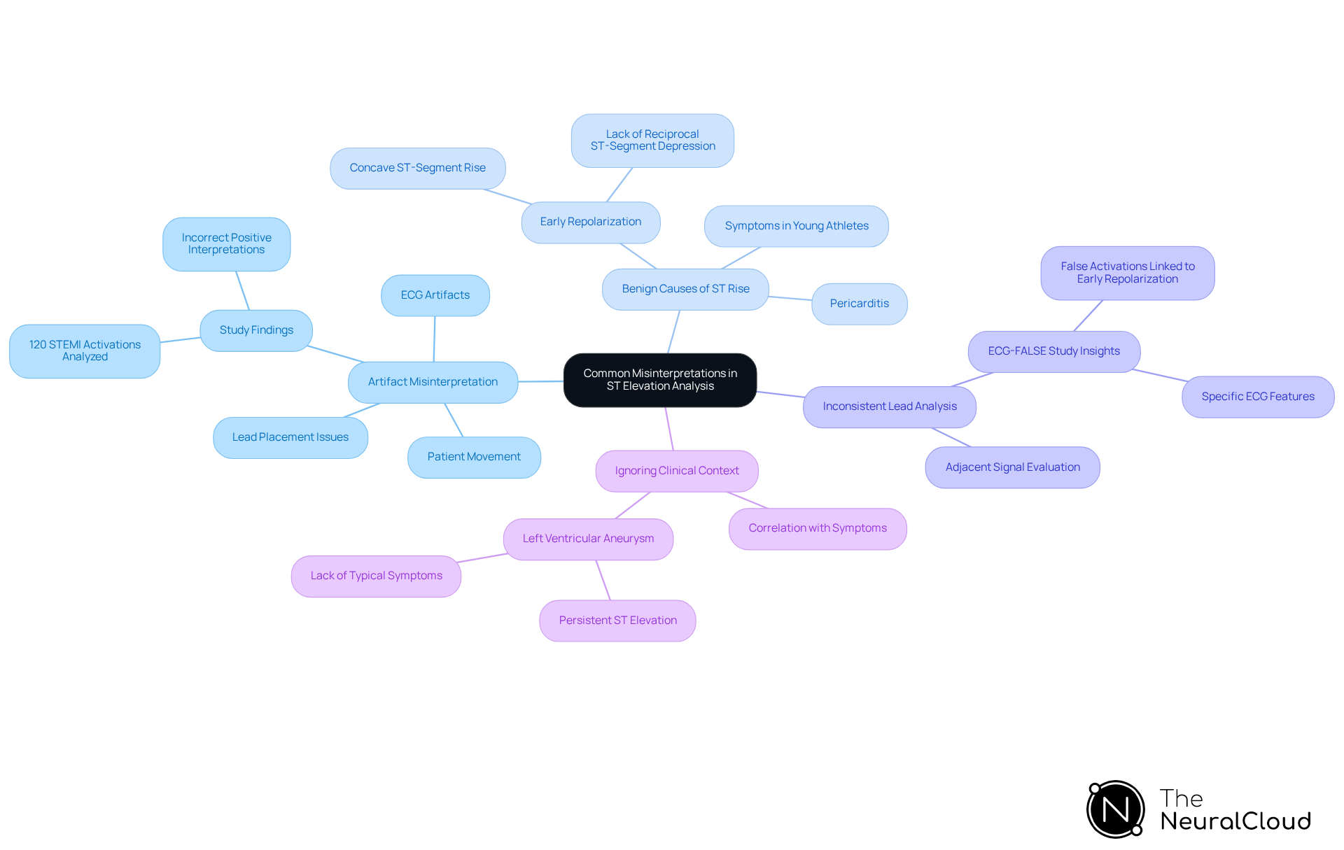
- Artifact Misinterpretation: ECG artifacts can mimic ST elevation. Always check for lead placement issues or patient movement that may distort the signal. In a study examining 120 STEMI activations, incorrect positive interpretations were mainly linked to ECG artifacts and non-ischemic reasons for ST increase.
- Benign Causes of ST Rise: Conditions such as early repolarization or pericarditis can cause ST rise without myocardial infarction. Familiarize yourself with these benign patterns to avoid unnecessary interventions. Early repolarization, frequently observed in young athletes, is marked by concave ST-segment rise and the lack of reciprocal ST-segment depression, which can easily be confused with acute myocardial infarction.
- Inconsistent Lead Analysis: Failing to analyze all relevant prospects can result in missed diagnoses. Always evaluate adjacent signals to confirm ST elevation. The ECG-FALSE study highlighted that specific ECG features, such as the presence of early repolarization, were associated with false activations, emphasizing the importance of thorough lead analysis.
- Ignoring Clinical Context: Relying solely on ECG findings without considering the individual's clinical presentation can result in misdiagnosis. Always correlate ECG results with the individual's symptoms and history. In cases of left ventricular aneurysm, persistent ST elevation may occur without the typical symptoms of myocardial infarction, necessitating careful clinical correlation.
By understanding these pitfalls, healthcare professionals can significantly enhance their diagnostic accuracy and improve patient outcomes.
Conclusion
Navigating the complexities of normal sinus rhythm with ST elevation presents significant challenges for healthcare professionals, particularly in the Canadian context where timely and accurate diagnoses can significantly impact patient outcomes. Understanding these characteristics is essential for accurate diagnoses, equipping clinicians with the necessary tools to make informed decisions in critical situations.
In this tutorial, we’ve explored key insights that can help you improve your ECG interpretation skills, including the defining features of normal sinus rhythm, the implications of ST elevation, and a systematic approach to ECG analysis. We emphasized the importance of recognizing common misinterpretations that can lead to diagnostic errors. By utilizing advanced tools like MaxYield™ from Neural Cloud Solutions Inc., clinicians can transform their ECG analysis, leading to more reliable patient assessments.
In conclusion, the ability to accurately identify normal sinus rhythm with ST elevation is not just a technical skill; it is a vital component of cardiovascular care that can save lives. Healthcare professionals are encouraged to continually refine their ECG interpretation skills and leverage innovative technologies to improve diagnostic accuracy. Embracing advanced technologies like MaxYield™ not only enhances diagnostic precision but also elevates the standard of cardiovascular care in Canada.
Frequently Asked Questions
What is Normal Sinus Rhythm (NSR)?
Normal Sinus Rhythm (NSR) is characterized by electrical impulses originating from the sinoatrial (SA) node, resulting in a consistent heart rhythm.
What are the key characteristics of Normal Sinus Rhythm?
The key characteristics of NSR include:
- Heart Rate: Typically ranges from 60 to 100 beats per minute (bpm).
- P Waves: Each P wave precedes a QRS complex, must be round, identical in shape, and present before every QRS complex in a 1:1 ratio.
- PR Interval: Consistent interval between the P wave and the QRS complex, usually between 0.12 and 0.20 seconds.
- QRS Complex: Narrow and uniform, typically less than 0.12 seconds in duration.
- Rhythm Regularity: Consistent R-R intervals with less than 0.16-second variation between successive P waves.
Why is understanding NSR important for healthcare professionals?
Understanding NSR is essential for healthcare professionals to accurately distinguish it from other cardiac rhythms, particularly in the context of ST elevation analysis, which can significantly impact clinical outcomes.
How does the MaxYield™ platform assist in interpreting Normal Sinus Rhythm?
The MaxYield™ platform from Neural Cloud Solutions Inc. addresses inaccuracies in ECG data by providing hospital-grade ECG accuracy from consumer-grade devices, ensuring clean data and transforming noisy recordings into detailed insights.
What are the benefits of using MaxYield™ for ECG interpretation?
MaxYield™ delivers beat-by-beat analysis of 200,000 heartbeats in less than 5 minutes, reduces error variance in downstream models, and supports confident clinical decisions, ultimately leading to better patient outcomes.
Who endorses the MaxYield™ platform and what do they say about it?
Experts such as Dr. Alan Rabinowitz, Dr. Brett Heilbron, and Dr. Marc W. Deyell endorse MaxYield™, emphasizing its precision and ability to significantly reduce noise in ECG data, rivaling human interpretation.
What is the regulatory status of MaxYield™?
MaxYield™ has FDA Class II SaMD clearance-pending status and audit-ready documentation supporting GxP inspections, making it a reliable tool for clinicians and researchers.
List of Sources
- Define Normal Sinus Rhythm and Its Characteristics
- Normal Sinus Rhythm (https://litfl.com/normal-sinus-rhythm-ecg-library)
- Sinus rhythm - Wikipedia (https://en.wikipedia.org/wiki/Sinus_rhythm)
- Sinus Rhythms Explained: A Comprehensive Guide to ECG Interpretation (https://aclscertification.org/rhythms-originating-from-the-sinus-node)
- Explain ST Elevation: Clinical Significance and Interpretation
- STEMI ECG Criteria: ST-Elevation Thresholds (https://powerfulmedical.com/blog/stemi-ecg-criteria)
- academic.oup.com (https://academic.oup.com/eurjpc/article/31/Supplement_1/zwae175.082/7692567)
- Acute ST-Segment Elevation Myocardial Infarction (STEMI) - StatPearls - NCBI Bookshelf (https://ncbi.nlm.nih.gov/books/NBK532281)
- Guide Through ECG Analysis: Steps to Identify Normal Sinus Rhythm with ST Elevation
- Cause of ST-segment elevation on electrocardiogram - PMC (https://pmc.ncbi.nlm.nih.gov/articles/PMC9578122)
- Accuracy of ECG chest electrode placements by paramedics: an observational study - PMC (https://pmc.ncbi.nlm.nih.gov/articles/PMC8312365)
- ECG Rhythm Evaluation (https://litfl.com/ecg-rhythm-evaluation)
- Steps to interpret an electrocardiogram for healthcare professionals (https://hvt-journal.com/articles/art254)
- Identify Common Misinterpretations in ST Elevation Analysis
- STEMI Mimics: ECG Impostors of Heart Attack (https://powerfulmedical.com/blog/stemi-mimics)
- Causes of Prehospital Misinterpretations of ST Elevation Myocardial Infarction (https://tandfonline.com/doi/full/10.1080/10903127.2016.1247200)
- Technical Factors Involved in False Positive ECG STEMI Diagnoses (https://dicardiology.com/article/technical-factors-involved-false-positive-ecg-stemi-diagnoses)