Introduction
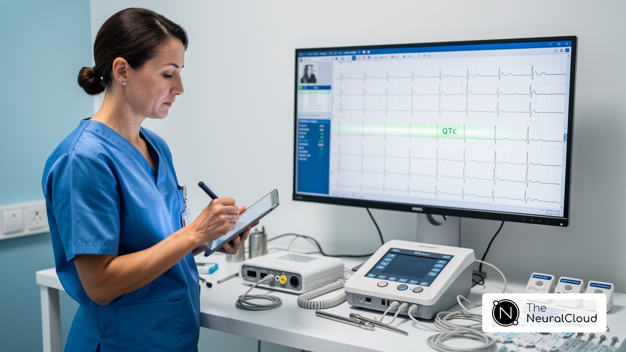
Clinicians often struggle with the complexities of RCA infarct ECG interpretation, which is crucial for effective patient care. This guide offers a streamlined approach to mastering the interpretation of RCA infarct ECGs, focusing on key features and systematic techniques that enhance diagnostic accuracy.
Clinicians often face challenges in distinguishing RCA infarcts from other conditions due to similar symptoms. This misinterpretation can result in delayed treatment and adverse patient outcomes, raising the question of how healthcare providers can ensure accurate identification of RCA infarcts.
Understand RCA Infarct Basics
The right coronary artery (RCA) is vital for supplying blood to the heart, and its occlusion can lead to significant clinical challenges. An RCA infarct ECG typically results from occlusion, which leads to ischemia in the areas it perfuses. Grasping the anatomy and physiology of the RCA is crucial for accurate ECG interpretation, particularly with advanced tools like MaxYield™. Key points include:
- Anatomy: The RCA branches into the right marginal artery and the posterior descending artery, which supply the inferior wall and parts of the septum. This anatomical knowledge is vital for identifying the affected regions during an infarct.
- Physiological Impact: Occlusion of the RCA can result in inferior wall myocardial infarction (MI), often presenting with symptoms such as bradycardia or AV block due to its supply to the sinoatrial (SA) and atrioventricular (AV) nodes. Approximately 40% of inferior wall MIs also involve the right ventricle, complicating clinical outcomes and associated with worse prognoses.
Clinical relevance involves recognizing the RCA infarct ECG's role, which is crucial for understanding the implications of ST-segment elevation in inferior leads (II, III, aVF) during an infarct. With a mortality rate between 2% and 9% for inferior wall MIs, timely diagnosis and intervention are essential to prevent serious complications. A simplified risk index has shown a prevalence of RCA involvement exceeding 90% among patients with scores greater than two, highlighting the RCA's significance in acute myocardial infarction scenarios. As noted, previously published criteria for identifying RCA involvement appear inadequate, making the recognition of RCA's role even more critical.
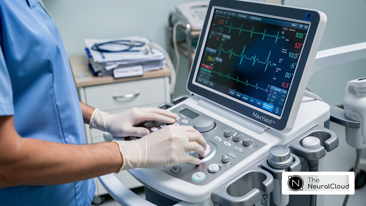
In this context, the system enhances ECG analysis by mapping ECG signals through noise, isolating and labeling key features in every heartbeat. Its advanced noise filtering capabilities allow for the identification of critical data even in recordings with high levels of noise and artifact, ensuring that previously obscured sections of lengthy Holter and patch monitor recordings can be salvaged. This automated analysis not only improves diagnostic yield but also supports confident clinical decision-making, making it an invaluable tool for health tech developers and clinicians alike. As Dr. Alan Rabinowitz observes, 'This system rivals human interpretation in precision, particularly in challenging cases like RCA infarct ECG.' Furthermore, with FDA Class II SaMD clearance-pending status, Neural Cloud Solutions Inc. ensures that their product meets rigorous regulatory standards, providing audit-ready documentation for GxP inspections. For those interested in exploring MaxYield™ further, Neural Cloud Solutions Inc. invites you to contact them for free trials or personalized walkthroughs.

Identify Key ECG Features of RCA Infarcts
Accurate interpretation of RCA infarct ECG can be challenging due to overlapping features and variations in presentation. When interpreting an ECG for RCA infarcts, focus on the following key features:
- ST Segment Elevation: Look for ST-segment elevation in leads II, III, and aVF, which indicates inferior wall ischemia. Notably, the elevation in channel III is usually higher than in channel II (ST III > ST II).
- Reciprocal Changes: Evaluate for ST-segment depression in positions I and aVL, which may suggest reciprocal changes linked to inferior myocardial infarctions (MIs).
- T-Wave Changes: Initially, hyperacute T-waves may be present, transitioning to T-wave inversions as the infarct evolves.
- Q Waves: The emergence of Q waves in the inferior electrodes may occur over time, indicating a more established infarct.
Recent studies highlight that up to 20% of patients with inferior STEMI may develop second- or third-degree AV block, emphasizing the importance of thorough ECG analysis. In a comprehensive review of 205,017 cases of myocardial infarction in Canada, marked ST elevation in channels II, III, and aVF was consistently observed, reinforcing the diagnostic significance of these features. Moreover, case studies show that hyperacute inferior STEMI appears with hyperacute T waves in positions II, III, and aVF, along with ST elevation and reduction of R wave height, which is indicative of an RCA infarct ECG confirming RCA occlusion.
Recognizing these features is crucial for timely intervention, ultimately improving patient outcomes in cases of RCA infarct ECG.

Interpret RCA Infarct ECG Readings
Interpreting the RCA infarct ECG readings presents unique challenges that require a systematic approach. To effectively interpret these readings, follow these essential steps:
- Evaluate the Signals: Begin by analyzing signals II, III, and aVF for ST-segment elevation. Ensure that electrode III shows greater elevation than electrode II, as this indicates RCA infarct ECG.
- Look for Reciprocal Changes: Examine electrodes I and aVL for ST-segment depression, which supports the diagnosis of an inferior myocardial infarction (MI). This reciprocal change is crucial for confirming the RCA infarct ECG occlusion.
- Evaluate T-Wave Morphology: Pay attention to any hyperacute T-waves, which may signal early ischemia. As the infarct evolves, T-wave inversions may appear, further indicating myocardial distress.
- Monitor for Q Waves: Over time, the appearance of Q waves in the inferior electrodes signifies a completed infarct, serving as a critical marker of myocardial necrosis. A pathological Q wave is defined as being more than 0.04 seconds in duration and must exceed 25% of the size of the following R waves in that lead, except for leads III and aVR.
- Consider Clinical Context: Always correlate ECG findings with the patient's clinical presentation, including symptoms and medical history, to confirm the diagnosis. This holistic approach ensures accurate interpretation and effective patient management.
Following these guidelines helps healthcare professionals improve diagnostic accuracy and enhance patient outcomes in myocardial infarction cases. Neural Cloud Solutions Inc.'s advanced platform features sophisticated noise filtering and unique wave recognition, greatly assisting in isolating essential data from noisy recordings. The user manual offers practical guidance on utilizing the product effectively in clinical settings. As Dr. Alan Rabinowitz notes, "MaxYield™ rivals human interpretation in precision, making it an invaluable tool for healthcare professionals." This ECG analysis solution automates labeling and adapts to physiological variability, helping clinicians tackle ECG interpretation challenges. With the right tools, healthcare professionals can navigate these complexities and improve patient care.

Troubleshoot Common Interpretation Challenges
Interpreting RCA infarct ECGs presents several challenges that can significantly impact patient care:
- Lead Misplacement: Accurate lead placement is crucial, as misplacement can lead to significant misinterpretations of ST-segment changes. Research shows that misplacement of limb electrodes can change diagnoses in about 25% of instances, possibly resulting in unnecessary procedures. This technology uses advanced noise filtering and unique wave recognition to reduce the effects of lead misplacement, delivering clearer ECG signals for analysis.
- Artifact Interference: Artifacts can mimic ST-segment elevation or depression, complicating the interpretation of ECGs. It is essential to confirm findings with a repeat ECG if artifacts are suspected, as they can lead to misdiagnoses and unnecessary stress testing. The 'CAPS SCAN' method can effectively identify such artifacts. The system excels in isolating ECG waves from recordings with high levels of noise and artifact, ensuring that clinicians can focus on accurate interpretations.
- Variability in Presentation: Patients may not always exhibit classic signs of RCA infarcts. Subtle changes or atypical presentations are particularly common in elderly patients or those with comorbidities, necessitating a careful review of the ECG in conjunction with clinical context. This product adapts with each use, improving diagnostic yield and accuracy over time, which is essential for these complex cases.
- Differentiating from Other Conditions: Distinguishing RCA infarcts from conditions such as pericarditis or left circumflex artery occlusion is critical, as these can present similarly on an ECG. Utilizing clinical context and additional diagnostic tests can aid in making accurate interpretations. Expert insights from Dr. Alan Rabinowitz and Dr. Brett Heilbron emphasize that MaxYield™ rivals human interpretation in precision, making it an invaluable tool in differentiating these conditions.
In a study assessing 9,424 adult ECGs, it was discovered that poor R-wave progression (PRWP) and reversed R-wave progression (RRWP) were common misinterpretations caused by electrode mispositioning, with prevalence rates varying from 15% to 42%. This highlights the need for improved training and adherence to standardized procedures. Misinterpretation due to lead misplacement can lead to unnecessary cardiology consultations and stress tests, contributing to an estimated cost burden of approximately $3.2 billion annually to the healthcare system due to ECG lead mispositioning. Integrating this system into their workflow helps healthcare providers cut costs and improve the accuracy of interpretations of RCA infarct ECG. Additionally, MaxYield™ is currently pending FDA Class II SaMD clearance, ensuring compliance with regulatory standards. For a more comprehensive analysis, Insight360 can be utilized as a customizable visualization and reporting tool that transforms MaxYield’s clean ECG data into interactive dashboards and clinical-ready reports.

Conclusion
Timely and accurate diagnosis of RCA infarct ECGs is crucial for healthcare professionals, as it can dramatically influence patient outcomes. This article presents a systematic approach to mastering this critical skill, emphasizing the importance of recognizing key features such as:
- ST-segment elevation
- Reciprocal changes
- T-wave morphology
- The presence of Q waves
By following these steps, clinicians can enhance their diagnostic accuracy and ensure effective patient management.
Key insights discussed include:
- The anatomical and physiological relevance of the right coronary artery
- The common challenges faced during interpretation
- The advanced tools available, such as MaxYield™ from Neural Cloud Solutions Inc.
These tools not only improve the clarity of ECG readings but also assist in overcoming obstacles like lead misplacement and artifact interference. The integration of technology in ECG analysis is a game-changer, enabling healthcare providers to deliver better care while reducing the risk of misdiagnosis.
Mastering RCA infarct ECG interpretation goes beyond technical knowledge; it’s about enhancing patient care and outcomes. Clinicians should take advantage of advanced tools and stay informed about the latest techniques to navigate the complexities of ECG interpretation effectively. By doing so, they can ensure that they are well-equipped to handle the challenges presented by RCA infarcts, ultimately leading to improved health outcomes for their patients.
Frequently Asked Questions
What is the significance of the right coronary artery (RCA) in heart health?
The RCA is vital for supplying blood to the heart, and its occlusion can lead to significant clinical challenges, including inferior wall myocardial infarction (MI).
What are the anatomical branches of the RCA?
The RCA branches into the right marginal artery and the posterior descending artery, which supply the inferior wall and parts of the septum.
What physiological impacts can result from RCA occlusion?
Occlusion of the RCA can lead to inferior wall MI, often presenting with symptoms such as bradycardia or AV block due to its supply to the sinoatrial (SA) and atrioventricular (AV) nodes.
How common is right ventricle involvement in inferior wall MIs?
Approximately 40% of inferior wall MIs also involve the right ventricle, complicating clinical outcomes and associated with worse prognoses.
Why is recognizing RCA infarct ECG important?
Recognizing the RCA infarct ECG is crucial for understanding the implications of ST-segment elevation in inferior leads (II, III, aVF) during an infarct, as timely diagnosis and intervention can prevent serious complications.
What is the mortality rate for inferior wall MIs?
The mortality rate for inferior wall MIs ranges between 2% and 9%.
What does the simplified risk index indicate about RCA involvement?
The simplified risk index shows that RCA involvement exceeds 90% among patients with scores greater than two, highlighting its significance in acute myocardial infarction scenarios.
How does MaxYield™ enhance ECG analysis?
MaxYield™ enhances ECG analysis by mapping ECG signals through noise, isolating and labeling key features in every heartbeat, and improving diagnostic yield even in recordings with high levels of noise and artifact.
What is the regulatory status of Neural Cloud Solutions Inc.'s product?
Neural Cloud Solutions Inc. is pursuing FDA Class II SaMD clearance-pending status, ensuring that their product meets rigorous regulatory standards and provides audit-ready documentation for GxP inspections.
How can interested parties explore MaxYield™ further?
Interested parties can contact Neural Cloud Solutions Inc. for free trials or personalized walkthroughs of MaxYield™.
List of Sources
- Understand RCA Infarct Basics
- Electrocardiographic identification of the culprit coronary artery in inferior wall ST elevation myocardial infarction (https://sciencedirect.com/science/article/abs/pii/S0828282X10703925)
- AI-based ECG interpretation outperforms standard diagnosis of occlusive myocardial infarction (https://news-medical.net/news/20260323/AI-based-ECG-interpretation-outperforms-standard-diagnosis-of-occlusive-myocardial-infarction.aspx)
- Inferior Myocardial Infarction - StatPearls - NCBI Bookshelf (https://ncbi.nlm.nih.gov/books/NBK470572)
- Identify Key ECG Features of RCA Infarcts
- Inferior STEMI (https://litfl.com/inferior-stemi-ecg-library)
- A narrative review of electrocardiogram manifestation of myocardial infarction with non-obstructive coronary arteries (MINOCA) (https://jtd.amegroups.org/article/view/109625/html)
- Temporal Trends in Incidence and Outcomes of Acute Myocardial Infarction in a Canadian Province: A 23-Year Analysis (https://sciencedirect.com/science/article/pii/S2589790X26003215)
- Interpret RCA Infarct ECG Readings
- ECGs in Acute Myocardial Infarction | ACLS Training (https://aclsmedicaltraining.com/ecg-in-acute-myocardial-infarction)
- Inferior STEMI (https://litfl.com/inferior-stemi-ecg-library)
- Electrocardiographic identification of the culprit coronary artery in inferior wall ST elevation myocardial infarction (https://sciencedirect.com/science/article/abs/pii/S0828282X10703925)
- AI-based ECG interpretation outperforms standard diagnosis of occlusive myocardial infarction (https://news-medical.net/news/20260323/AI-based-ECG-interpretation-outperforms-standard-diagnosis-of-occlusive-myocardial-infarction.aspx)
- Troubleshoot Common Interpretation Challenges
- ECG Lead Misplacement: Looking at Common Issues (https://gehealthcare.com/en-gb/insights/article/ecg-lead-misplacement-looking-at-common-issues?srsltid=AfmBOoq4uoWgLCHPJh5cLR4WJOVvfv6qS_m3nm1Z1maHKqP8BQV_rBYK)
- Misdiagnosis from ECG Lead Misplacement, Artifact and Lead Reversal | ECG Cases (https://emergencymedicinecases.com/ecg-cases-lead-misplacement-artifact-lead-reversal)
- Precordial ECG Lead Mispositioning: Its Incidence and Estimated Cost to Healthcare - PMC (https://pmc.ncbi.nlm.nih.gov/articles/PMC7343296)
- ECG Lead Misplacement -Fool Me Once Shame on You, Fool Me Twice Shame on Me (https://austinpublishinggroup.com/emergency-medicine/fulltext/aem-v2-id1025.php)
- ECGs in Acute Myocardial Infarction | ACLS Training (https://aclsmedicaltraining.com/ecg-in-acute-myocardial-infarction)