Introduction
Navigating the complexities of ECG analysis presents significant challenges for clinicians, especially when diagnosing acute coronary syndromes like STEMI. Let’s explore the critical role of ST elevation leads, focusing on how to interpret them, their clinical significance, and the latest innovations in analysis technology. Yet, with various conditions mimicking ST elevation, clinicians must navigate these complexities to ensure accurate diagnoses and timely interventions that could save lives.
Clarify the Basics of ST Elevation Leads
The ST segment is a critical component of ECG analysis, representing the interval between ventricular depolarization and repolarization. It is characterized as the flat, isoelectric section of the ECG that lies between the end of the S wave (the J point) and the onset of the T wave. Recognizing the importance of this segment is crucial, as any increase can signify serious heart issues, including myocardial ischemia or infarction.
In ST elevation leads, an ST increase is characterized by an upward shift above the baseline, necessitating a measurement of at least 1 mm in two adjacent leads. This elevation is most commonly linked to ST elevation leads in ST-Elevation Myocardial Infarction, which represents 30-33% of all acute coronary syndromes (ACS). Comprehending the typical morphology of the ST segment and its variations across different leads is essential for precise diagnosis and efficient treatment planning.
Despite the initial non-diagnostic ECG, timely interpretation is critical for effective treatment. Statistics indicate that 11.0% of individuals diagnosed with a heart attack initially show a non-diagnostic ECG, yet 72.4% receive a diagnostic ECG within 90 minutes. This underscores the need for advanced ECG analysis tools to ensure rapid and accurate diagnosis. Furthermore, the documented mortality rate for ST elevation myocardial infarction was between 5.0-8.0% in 2006, emphasizing the crucial importance of identifying ST changes swiftly.
Case studies demonstrate the function of the 12-lead ECG in identifying ST elevation leads in myocardial infarction, where particular ST changes and T-wave inversions are crucial signs. For example, a coronary angiogram may be conducted after the detection of elevated ST intervals to identify blockages, enabling focused treatment and enhancing patient results.
In summary, the rise of the ST segment acts as a crucial sign in ECG analysis, particularly in the context where ST elevation leads to myocardial infarction, where swift diagnosis and intervention can greatly influence patient outcomes. With MaxYield™, health tech developers can leverage advanced ECG analysis capabilities, including the integration of Insight360 for customizable visualization and reporting, to enhance clinical decision-making and improve patient care.

Analyze ECG Patterns of ST Elevation in STEMI
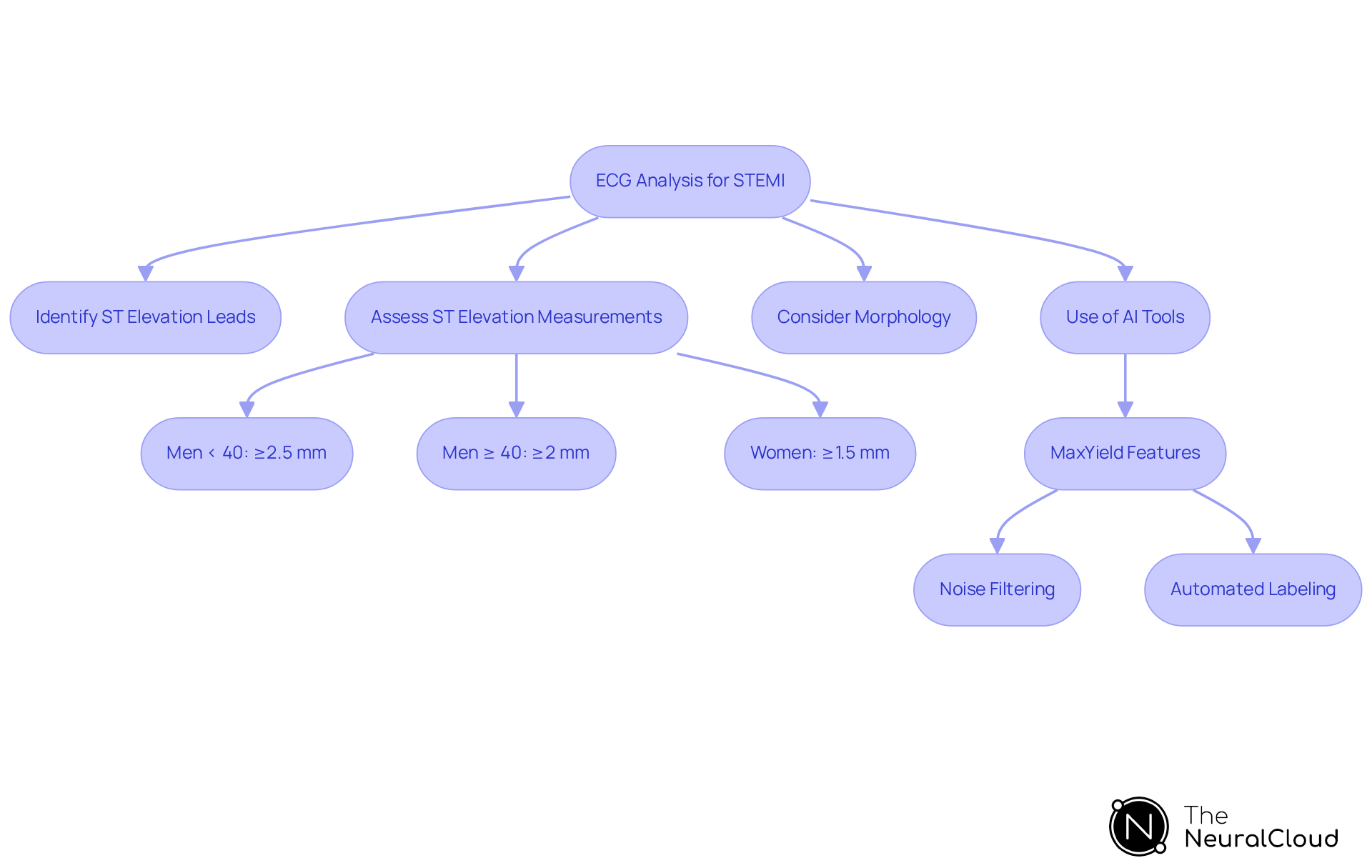
In the realm of STEMI diagnosis, recognizing that [ST elevation leads](https://saem.org/about-saem/academies-interest-groups-affiliates2/cdem/for-students/online-education/m3-curriculum/group-electrocardiogram-(ecg)-rhythm-recognition/stemi) is critical for effective patient management. The ECG typically displays distinct patterns of ST elevation leads, which are essential for diagnosis. Clinicians should identify ST elevation leads showing an ST rise of at least 1 mm in two or more contiguous leads, a hallmark of this condition. The ST elevation leads, especially V2 and V3, are crucial for identifying increases that suggest anterior wall involvement. For men under 40 years, an increase of ≥2.5 mm in these leads is required, while men aged 40 and older need ≥2 mm, and women of any age require ≥1.5 mm for a definitive diagnosis.
The morphology of the ST segment also provides essential diagnostic clues. A convex or 'tombstone' appearance is often linked to acute STEMI, indicating a more severe condition, while a concave rise may suggest alternative diagnoses. It's crucial for healthcare professionals to understand these patterns. This knowledge helps them differentiate between types of myocardial infarctions and start the right treatment quickly. For example, the Sgarbossa criteria can assist in evaluating ST rise in patients with left bundle branch block (LBBB), where conventional definitions may not be relevant.
Recent case studies highlight the importance of timely ECG interpretation. Delays in ECG interpretation can lead to worsened patient outcomes. For instance, a study demonstrated that quick identification of ST elevation leads to a significant decrease in treatment delays, enhancing patient outcomes. Moreover, AI-powered technologies such as Neural Cloud Solutions' MaxYield can decrease the time to diagnosis by 3 hours, improving the sensitivity of ECG analysis and enabling faster and more precise identification of heart attacks. MaxYield offers advanced noise filtering and automated labeling. These features turn noisy recordings into clear insights, helping healthcare professionals respond effectively to critical conditions. With the ability to analyze 200,000 heartbeats in less than 5 minutes, MaxYield ensures that clinicians have access to detailed, beat-by-beat analysis, which is crucial given that every minute of delay in treatment increases myocardial damage. This emphasizes the goal of achieving a Door to Needle time for administering thrombolytics of less than 30 minutes. By mastering the analysis of ST elevation leads with tools like MaxYield, healthcare professionals can enhance their clinical decision-making and improve patient outcomes. With tools like MaxYield, healthcare professionals can not only enhance their diagnostic accuracy but also save lives through timely intervention.
Differentiate ST Elevation from Other Cardiac Conditions
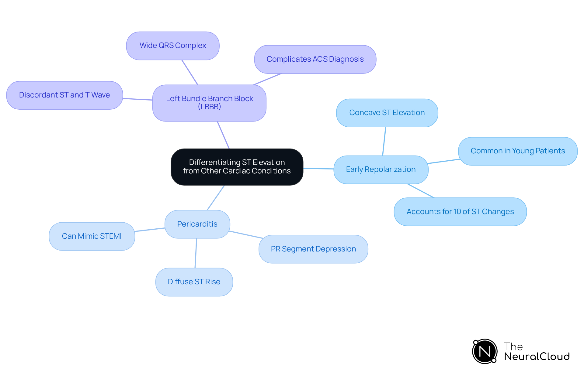
ST elevation leads are a critical marker in ECG analysis, but they can indicate various conditions beyond STEMI. Clinicians must be adept at distinguishing these presentations to avoid misdiagnosis and unnecessary interventions. Key conditions that may present with ST segment elevation include:
- Early Repolarization: This benign variant typically shows a concave ST elevation, predominantly in younger patients without cardiac symptoms. It represents roughly 10% of ST changes in emergency department patients experiencing chest pain.
- Pericarditis: Characterized by diffuse ST rise across multiple leads, pericarditis often presents with accompanying PR segment depression. This pattern can be mistaken for STEMI, particularly in cases of focal pericarditis following myocardial infarction or surgery.
- Left Bundle Branch Block (LBBB): While LBBB can show ST rise, it is typically linked with a wide QRS complex. The ST portion and T wave are often discordant to the primary direction of the QRS complex, complicating the diagnostic process for acute coronary syndrome (ACS).
Recognizing these differences is crucial for making accurate diagnoses and providing effective treatment. Research shows that 60%-80% of individuals with ST elevation leads do not have STEMI, emphasizing the commonality of STEMI imitators. Misinterpretation can lead to unnecessary procedures, underscoring the importance of accurate ECG analysis. Utilizing advanced tools like MaxYield™ can significantly reduce the risk of misdiagnosis and improve patient care outcomes. Additionally, the Insight360 tool transforms MaxYield’s clean ECG data into interactive dashboards and clinical-ready reports, further improving the accuracy and efficiency of ECG interpretations.
Leverage Advanced Technologies for Enhanced ECG Analysis
Noisy ECG recordings often lead to misinterpretations, complicating accurate diagnoses in cardiology. The integration of advanced technologies, particularly AI and machine learning, into ECG analysis marks a pivotal evolution in this field. Solutions like Neural Cloud Solutions' MaxYield significantly enhance the accuracy of detecting ST elevation leads by effectively isolating and cleaning noisy signals, which facilitates clearer interpretations.
With advanced noise filtering and distinct wave recognition, MaxYield™ identifies and labels critical data effectively, even in recordings with high levels of noise and artifact. This capability rapidly isolates ECG waves from recordings affected by baseline wander, movement, and muscle artifact. It salvages previously obscured sections of lengthy Holter, 1-Lead, and patch monitor recordings. By transforming noisy recordings into detailed insights, MaxYield™ delivers beat-by-beat analysis, outputting an analysis of 200,000 heartbeats in less than 5 minutes.
AI algorithms process extensive ECG datasets swiftly, identifying intricate patterns that may elude human observation. For example, convolutional neural networks (CNNs) can be trained to detect subtle variations in ST segment morphology, which can improve the identification of ST elevation leads and enhance diagnostic precision. Recent studies have demonstrated that AI models can outperform traditional clinical tests, identifying high-risk patients for sudden cardiac death with a 7% annual rate, compared to 4.6% through conventional methods. This capability not only streamlines workflows for healthcare professionals but also accelerates the accuracy of ECG data analysis, ultimately enhancing patient care.
Moreover, MaxYield's continuous learning model evolves with each use, improving its diagnostic yield over time. Clinicians are encouraged to integrate MaxYield into their existing platforms to leverage its capabilities fully. Ziad Obermeyer, the study lead author, highlights the significant potential of AI in enhancing medical decision-making, especially given that sudden cardiac arrest leads to over 300,000 deaths annually in the U.S. By adopting MaxYield, clinicians can significantly enhance diagnostic accuracy and patient outcomes, ultimately transforming cardiovascular care.

Conclusion
Healthcare professionals face significant challenges in accurately analyzing ST elevation leads, which are crucial for effective cardiac care, particularly in the context of ST-Elevation Myocardial Infarction (STEMI). Recognizing the specific patterns of ST segment elevation is vital for accurate diagnosis and significantly impacts patient outcomes. By leveraging advanced technologies like MaxYield, healthcare professionals can enhance their diagnostic capabilities, ensuring timely and precise interventions that can save lives.
Throughout the article, key insights were shared regarding the critical role of ST elevation leads in ECG analysis. The importance of recognizing distinct patterns, differentiating ST elevation from other cardiac conditions, and utilizing innovative tools for enhanced analysis were emphasized. The integration of AI and machine learning into ECG interpretation represents a transformative shift, allowing for improved accuracy and efficiency in diagnosing conditions that can lead to serious health complications.
As cardiology advances, it's crucial for clinicians to stay updated with the latest tools. Embracing advanced ECG analysis tools is not just beneficial but necessary. Clinicians are encouraged to adopt solutions like MaxYield to refine their diagnostic processes and ultimately improve patient care. By mastering the intricacies of ST elevation leads and utilizing cutting-edge technology, healthcare professionals can ensure they are at the forefront of cardiac care, ready to respond effectively to the challenges presented by acute coronary syndromes.
Frequently Asked Questions
What is the ST segment in ECG analysis?
The ST segment is the interval between ventricular depolarization and repolarization, represented as the flat, isoelectric section of the ECG that lies between the end of the S wave (the J point) and the onset of the T wave.
Why is the ST segment important in diagnosing heart issues?
An increase in the ST segment can signify serious heart issues, including myocardial ischemia or infarction, making its recognition crucial for accurate diagnosis and treatment planning.
How is ST elevation defined in ECG leads?
ST elevation is characterized by an upward shift above the baseline, requiring a measurement of at least 1 mm in two adjacent leads.
What condition is most commonly associated with ST elevation leads?
ST elevation leads are most commonly linked to ST-Elevation Myocardial Infarction, which accounts for 30-33% of all acute coronary syndromes (ACS).
What is the significance of timely ECG interpretation?
Timely interpretation of ECGs is critical for effective treatment, as statistics show that 11.0% of individuals diagnosed with a heart attack initially present with a non-diagnostic ECG, yet 72.4% receive a diagnostic ECG within 90 minutes.
What was the documented mortality rate for ST elevation myocardial infarction in 2006?
The documented mortality rate for ST elevation myocardial infarction was between 5.0-8.0% in 2006.
How does a 12-lead ECG assist in identifying ST elevation leads?
A 12-lead ECG helps identify ST elevation leads in myocardial infarction by detecting specific ST changes and T-wave inversions, which are crucial signs for diagnosis.
What role does a coronary angiogram play after detecting elevated ST intervals?
A coronary angiogram may be conducted to identify blockages after detecting elevated ST intervals, enabling focused treatment and improving patient outcomes.
How can health tech developers enhance ECG analysis capabilities?
Health tech developers can leverage advanced ECG analysis capabilities, such as those offered by MaxYield™, including the integration of Insight360 for customizable visualization and reporting, to enhance clinical decision-making and improve patient care.
List of Sources
- Clarify the Basics of ST Elevation Leads
- What Is a STEMI? (https://my.clevelandclinic.org/health/diseases/22068-stemi-heart-attack)
- ST-segment Elevation Myocardial Infarction: Challenges in Diagnosis (https://uscjournal.com/articles/st-segment-elevation-myocardial-infarction-challenges-diagnosis?language_content_entity=en)
- STEMI: ST-segment Elevation Myocardial Infarction - The Cardiology Advisor (https://thecardiologyadvisor.com/ddi/stemi-st-elevation-myocardial-infarction)
- Analyze ECG Patterns of ST Elevation in STEMI
- STEMI ECG Criteria: ST-Elevation Thresholds (https://powerfulmedical.com/blog/stemi-ecg-criteria)
- STEMI - Electrocardiogram - M3 Curriculum | SAEM (https://saem.org/about-saem/academies-interest-groups-affiliates2/cdem/for-students/online-education/m3-curriculum/group-electrocardiogram-(ecg)-rhythm-recognition/stemi)
- What Is a STEMI? (https://my.clevelandclinic.org/health/diseases/22068-stemi-heart-attack)
- STEMI: ST-segment Elevation Myocardial Infarction - The Cardiology Advisor (https://thecardiologyadvisor.com/ddi/stemi-st-elevation-myocardial-infarction)
- Differentiate ST Elevation from Other Cardiac Conditions
- Checking your browser - reCAPTCHA (https://pmc.ncbi.nlm.nih.gov/articles/PMC4209433)
- ST‑segment elevation myocardial infarction mimics: The differential diagnosis of nonacute coronary syndrome causes of ST‑segment/T‑wave abnormalities in the chest pain patient | 2024, Volume 24, Issue 4 | Turkish Journal of Emergency Medicine (https://turkjemergmed.com/full-text/895)
- STEMI Mimics: ECG Impostors of Heart Attack (https://powerfulmedical.com/blog/stemi-mimics)
- ST Segment - StatPearls - NCBI Bookshelf (https://ncbi.nlm.nih.gov/books/NBK459364)
- Leverage Advanced Technologies for Enhanced ECG Analysis
- With AI, researchers discover new way to detect sudden cardiac death risk - Berkeley News (https://news.berkeley.edu/2026/06/24/with-ai-researchers-discover-new-way-to-detect-sudden-cardiac-death-risk)
- A sweeping new AI to detect heart conditions is coming to OpenEvidence (https://statnews.com/2026/06/23/pathway-labs-echonext-ai-tool-heart-disease-detection)
- AI-powered electrocardiogram detects early signs of heart failure (https://utsouthwestern.edu/newsroom/articles/year-2026/may-ai-powered-electrocardiogram.html)