Introduction
Understanding the heart's electrical activity is essential for effective diagnosis and treatment in cardiology. The 12-lead ECG serves as a cornerstone of this practice. By mastering the intricacies of electrode placement, healthcare professionals can significantly enhance their diagnostic capabilities. This leads to quicker and more accurate assessments of cardiac conditions. However, a challenge remains: how can one ensure precise placement amidst the complexities of human anatomy and technology?
This article explores essential steps and visual guides to navigate the nuances of 12-lead ECG placement. It aims to empower practitioners to avoid common pitfalls and improve patient outcomes.
Understand the Basics of 12-Lead ECG
A 12-lead ECG is a vital diagnostic tool that captures the heart's electrical activity, providing comprehensive insights into cardiac function. Each electrode—categorized into limb electrodes (I, II, III) and chest electrodes (V1-V6)—offers unique information essential for identifying various cardiac conditions. Understanding the anatomical relation of these electrodes corresponds to particular heart areas and is crucial for precise interpretation and diagnosis. For instance, the P wave reflects atrial depolarization, while the QRS complex indicates ventricular depolarization, both of which are critical for assessing heart health.
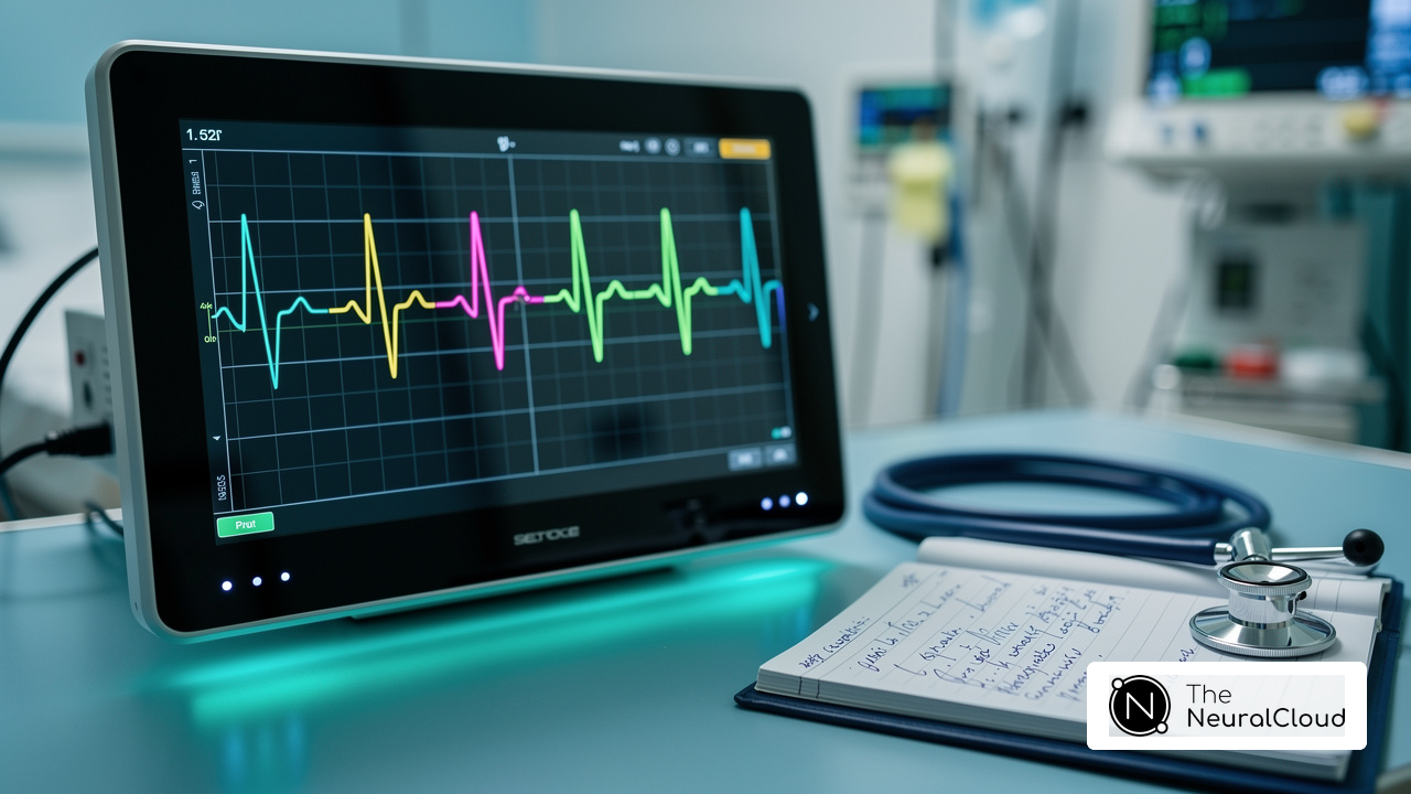
Neural Cloud Solutions' technology enhances this process by mapping ECG signals through noise, isolating and labeling key features in every heartbeat. This advanced system allows for the identification and labeling of critical data, even in recordings with high levels of noise and artifact. By rapidly isolating ECG waves from recordings affected by baseline wander, movement, and muscle artifact, MaxYield™ salvages previously obscured sections of lengthy Holter, 1-Lead, and patch monitor recordings. Specifically, MaxYield™ delivers beat-by-beat analysis, outputting heart rate, QRS complex, and T-wave onsets, offsets, and time-series intervals, addressing the challenges of noise reduction, and overcoming traditional limitations in ECG interpretation.
Case studies highlight the significance of technology in diagnosing conditions such as myocardial infarction and arrhythmias. For example, the Bradford District Care Foundation Trust reported a reduction in diagnosis time from several weeks to just a couple of days after implementing solutions like MaxYield™. This is crucial, as nearly 200 million ECGs are recorded annually worldwide, underscoring the widespread reliance on this tool in clinical practice.
Moreover, cardiologists emphasize the importance of comprehending the placement of electrodes for accurate diagnosis. Misplacement can lead to misdiagnosis, which may delay necessary treatment. As Lawrence Rosenthal noted, "A well-planned approach to 12-lead ECG interpretation will prevent the interpreter from missing crucial information." By familiarizing oneself with the heart's anatomy and the electrical impulses that govern its function, healthcare professionals can significantly enhance their diagnostic capabilities and improve patient outcomes, especially when leveraging advanced solutions like MaxYield™.

Recognize the Importance of Accurate Electrode Placement
Precise placement of sensors is essential for obtaining a clear and dependable ECG, leading to erroneous interpretations and potentially detrimental clinical decisions.
For instance, improper placement of electrodes can conceal crucial details about the heart's anterior wall, which is vital for diagnosing issues such as myocardial infarction. Research indicates that only 10% of healthcare professionals achieve perfect placement, highlighting the importance of accuracy, with frequent mistakes involving:
- V1 and V2 being set too high
- V5 and V6 too near the center
Such misplacements can result in misdiagnoses, such as false indications of right bundle branch block or ST-segment elevation.
A case study involving a 74-year-old patient illustrated that initial ECG readings with torso limb connections indicated acute inferior wall ischemia, which was later rectified by adjusting the connections to standard positions. This underscores the importance of proper placement during the procedure.
As specialists emphasize, accurate placement, illustrated in a diagram, is essential for precise diagnosis and treatment, highlighting the significance of continuous education and training for healthcare professionals to reduce positioning errors and improve patient safety.

Follow Step-by-Step Instructions for Proper Electrode Placement
- Prepare the Patient: Position the patient comfortably, ideally lying down, to facilitate relaxation during the procedure. Completely cleanse the skin at contact points to remove oils and dirt, ensuring optimal connection for precise readings. Utilizing technology enhances this process by improving efficiency, streamlining the preparation phase and mitigating issues related to electrode placement.
- Identify lead positions by familiarizing yourself with the electrode placement guide. Limb Leads: Attach electrodes to the right arm (RA), left arm (LA), right leg (RL), and left leg (LL). Note that limb lead sensors should not be placed on the wrists or ankles, as recommended by the American Heart Association. Chest Leads: Position V1 at the fourth intercostal space to the right of the sternum, V2 at the fourth intercostal space to the left, V3 between V2 and V4, V4 at the fifth intercostal space at the midclavicular line, V5 at the same horizontal level as V4 at the anterior axillary line, and V6 at the same level at the midaxillary line.
- Attach Sensors: Securely affix the sensors to the skin, ensuring good contact while avoiding bony areas or regions with excessive hair, which can interfere with signal quality. The platform enhances accuracy by providing stability and reducing the impact of motion artifacts.
- Connect Wires: Attach the wires from the ECG machine, verifying that each wire is linked to its assigned location.
- Check Connections: Prior to initiating the ECG, meticulously inspect and ensure that the electrodes are firmly placed and secure. This stage is essential, as incorrect positioning of the conductor can cause misinterpretation of outcomes, potentially influencing patient care. Furthermore, it is recommended to indicate the limitations for ECGs recorded with torso limb lead positions to inform clinicians of their limitations, as emphasized by professionals in the field. Leveraging best practices can help address challenges such as signal interference and patient movement, ensuring a smoother analysis process.

Troubleshoot Common Electrode Placement Issues
- Poor Connections: Noise or artifacts in the ECG often stem from improper sensor placement. To ensure optimal performance, confirm that terminals are securely attached and that the skin is clean. If needed, re-clean the skin to improve adhesion and reduce noise. Research indicates that using consistent types of electrodes and fresh gel significantly enhances signal quality. Correct placement can ensure ECG device precision nearly 95% of the time. The software from Neural Cloud Solutions aids in identifying and labeling issues, even in recordings with high noise levels, thereby improving overall accuracy.
- Inconsistent Readings: Variability in readings may arise when sensors are placed over bony areas or excessive hair. Adjusting the placement to soft areas of the skin is essential. Research shows that less than 20% of cardiologists accurately position V1 and V2 leads, which emphasizes the necessity of a training program for proper placement. MaxYield™'s AI-driven analysis helps mitigate these inconsistencies by providing feedback and enhancing diagnostic yield.
- Lead Misconnections: Abnormal ECG results can result from lead misconnections. It is crucial to double-check that each lead connects to the correct contact point, as even a minor mix-up can lead to misleading interpretations. Incorrect connections complicate ECG analysis and jeopardize patient safety. The continuous learning model of the device enhances accuracy by adapting with each use, effectively addressing potential misconnection issues.
- Patient Movement: Movement during recording can introduce artifacts that distort the ECG signal. Instructing the patient to remain still throughout the procedure is vital. If movement is unavoidable, consider utilizing a 3-lead ECG system, which can yield clearer signals and better readings, particularly in dynamic settings. For accurate readings, it is essential to follow the manufacturer's guidelines, which helps maintain device accuracy nearly 95% of the time. Furthermore, MaxYield™'s advanced noise filtering capabilities can recover previously obscured sections of recordings, making it a valuable asset in managing challenges.

Conclusion
Mastering the placement of the 12 lead ECG is crucial for healthcare professionals aiming to enhance diagnostic accuracy and improve patient outcomes. This guide underscores the importance of precise electrode positioning, as even slight misplacements can result in significant misdiagnoses and treatment delays. By grasping the complexities of the 12 lead ECG and the relationship between electrodes and heart anatomy, practitioners can interpret results accurately and efficiently.
The article presents key insights, including:
- The necessity of accurate electrode placement
- A step-by-step process for achieving this
- Strategies for troubleshooting common issues during ECG recordings
It highlights the integration of advanced technologies, such as Neural Cloud Solutions' MaxYield™ platform, which revolutionizes the ECG process. This platform offers enhanced noise filtering and automated annotations, streamlining ECG analysis and improving diagnostic clarity.
As the reliance on 12 lead ECGs grows in clinical practice, it is essential for healthcare professionals to prioritize proper training and continuous education in this vital area. By committing to excellence in electrode placement and utilizing advanced tools, practitioners can significantly reduce the risk of errors, leading to improved patient care and outcomes. Embracing these practices not only enhances individual diagnostic capabilities but also contributes to the overall advancement of cardiac health management.
Frequently Asked Questions
What is a 12-lead ECG and why is it important?
A 12-lead ECG is a vital diagnostic tool that captures the heart's electrical activity from twelve distinct perspectives, providing comprehensive insights into cardiac function. It is essential for identifying various cardiac conditions.
What are the different types of electrodes used in a 12-lead ECG?
The electrodes are categorized into limb electrodes (I, II, III) and chest electrodes (V1-V6), each offering unique information related to specific areas of the heart.
What do the P wave and QRS complex represent in a 12-lead ECG?
The P wave reflects atrial depolarization, while the QRS complex indicates ventricular depolarization. Both are critical for assessing heart health.
How does MaxYield™ enhance the analysis of ECG signals?
MaxYield™ enhances the analysis by mapping ECG signals through noise, isolating and labeling key features in every heartbeat, even in recordings with high levels of noise and artifact.
What specific capabilities does MaxYield™ provide in ECG analysis?
MaxYield™ delivers beat-by-beat analysis, outputting detailed insights on P-wave, QRS complex, and T-wave onsets, offsets, and time-series intervals, addressing challenges of physiological variability and signal artifacts.
How has the implementation of advanced ECG technologies like MaxYield™ impacted diagnosis times?
The Bradford District Care Foundation Trust reported a reduction in diagnosis time from several weeks to just a couple of days after implementing advanced ECG technologies.
Why is understanding the 12-lead ECG placement crucial for healthcare professionals?
Understanding the 12-lead ECG placement is crucial because misplacement can lead to misdiagnosis, potentially delaying necessary treatment.
What advice did Lawrence Rosenthal provide regarding 12-lead ECG interpretation?
Lawrence Rosenthal emphasized that a well-planned approach to 12-lead ECG interpretation will prevent the interpreter from missing crucial information.
How many ECGs are recorded annually worldwide, and what does this indicate?
Nearly 200 million ECGs are recorded annually worldwide, underscoring the widespread reliance on this tool in clinical practice.
How can healthcare professionals improve their diagnostic capabilities with respect to ECGs?
By familiarizing themselves with the heart's anatomy and the electrical impulses that govern its function, healthcare professionals can significantly enhance their diagnostic capabilities, especially when leveraging advanced solutions like MaxYield™.
List of Sources
- Understand the Basics of 12-Lead ECG
- Normal Electrocardiography (ECG) Intervals: Normal Electrocardiography Intervals (https://emedicine.medscape.com/article/2172196-overview)
- ecg-od.com (https://ecg-od.com/case_studies/bradford-district-care-foundation-use-of-ecg-on-demands-ai-assisted-12-lead-ecg-interpretation-service)
- Electrocardiogram - StatPearls - NCBI Bookshelf (https://ncbi.nlm.nih.gov/books/NBK549803)
- Electrocardiography - Wikipedia (https://en.wikipedia.org/wiki/Electrocardiography)
- mdpi.com (https://mdpi.com/2673-3846/2/4/505)
- Recognize the Importance of Accurate Electrode Placement
- Assessing the Accuracy of ECG Chest Electrode Placement by EMS and Clinical Personnel Using Two Evaluation Methods | International Journal of Paramedicine (https://internationaljournalofparamedicine.com/index.php/ijop/article/view/2897)
- researchgate.net (https://researchgate.net/publication/346113079_Artificial_Intelligence_Machine_Learning_and_Reasoning_in_Health_Informatics-Case_Studies)
- gehealthcare.com (https://gehealthcare.com/insights/article/ecg-lead-misplacement-looking-at-common-issues)
- ECG Electrode Placement: Risks, Tech, and Growing Demand in 2025- (https://intcomedical.com/news/info/What-You-Need-to-Know-About-ECG-Electrode-Placement-in-2025.html)
- pmc.ncbi.nlm.nih.gov (https://pmc.ncbi.nlm.nih.gov/articles/PMC8415387)
- Follow Step-by-Step Instructions for Proper Electrode Placement
- ECG Lead positioning (https://litfl.com/ecg-lead-positioning)
- pmc.ncbi.nlm.nih.gov (https://pmc.ncbi.nlm.nih.gov/articles/PMC8415387)
- ECG Electrode Placement: Risks, Tech, and Growing Demand in 2025- (https://intcomedical.com/news/info/What-You-Need-to-Know-About-ECG-Electrode-Placement-in-2025.html)
- jems.com (https://jems.com/patient-care/12-lead-ecgs-room-for-improvement)
- medicalsearch.com.au (https://medicalsearch.com.au/buying-guide/understanding-ecg-machines-and-electrocardiography/f/24929)
- Troubleshoot Common Electrode Placement Issues
- idrc-crdi.ca (https://idrc-crdi.ca/sites/default/files/openebooks/396-6)
- researchgate.net (https://researchgate.net/publication/346113079_Artificial_Intelligence_Machine_Learning_and_Reasoning_in_Health_Informatics-Case_Studies)
- ECG Electrode Placement: Risks, Tech, and Growing Demand in 2025- (https://intcomedical.com/news/info/What-You-Need-to-Know-About-ECG-Electrode-Placement-in-2025.html)