Introduction
Understanding the nuances of inferior myocardial infarction (IMI) is critical for healthcare professionals. Timely and accurate diagnosis can significantly impact patient outcomes. This article explores the challenges in ECG analysis related to IMI, particularly focusing on the essential steps for interpreting ECGs. Readers will gain insights into recognizing key features such as ST-segment elevation and T-wave inversions.
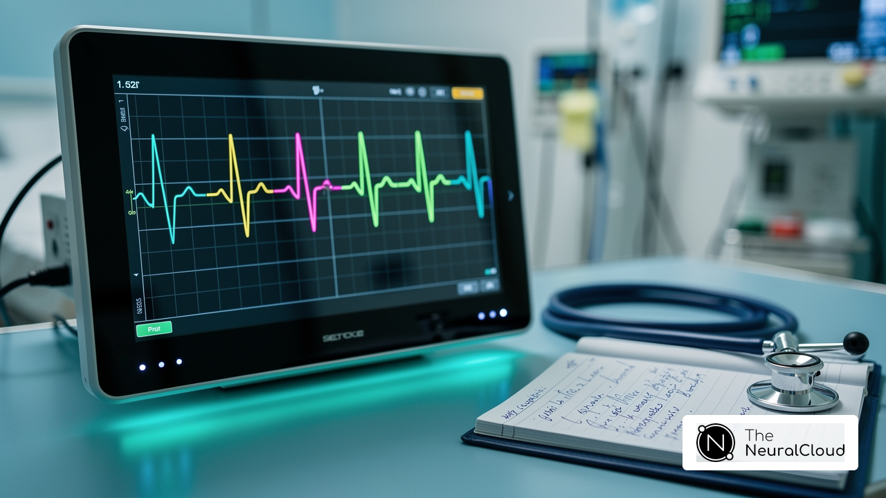
However, amidst the complexities of ECG analysis, common pitfalls can arise. Issues like lead misplacement and artifact interference can compromise diagnostic accuracy. To address these challenges, the MaxYield™ platform offers innovative features designed to enhance ECG interpretation. By utilizing advanced algorithms, it improves the accuracy of readings, ensuring healthcare professionals can make informed decisions.
The advantages of the MaxYield™ platform are clear. It not only streamlines the ECG analysis process but also provides real-time feedback, allowing for quicker diagnosis and treatment. This ultimately leads to better patient outcomes, as healthcare providers can respond promptly to critical situations.
In conclusion, understanding IMI and utilizing tools like MaxYield™ can significantly enhance diagnostic capabilities. By equipping healthcare professionals with the right knowledge and technology, we can improve patient care and outcomes.
Understand the Basics of Inferior Myocardial Infarction and ECG Interpretation
(IMI) occurs when blood flow to the heart is compromised, typically due to occlusion of the right coronary artery (RCA). Understanding the heart's structure and the specific electrodes associated with ECG interpretation is crucial for accurate diagnosis and treatment. The lower electrodes on a standard 12-electrode ECG - II, III, and aVF - provide essential information about the heart's electrical activity in this area.
Key Concepts:
- Definition: This condition is characterized by the death of heart muscle due to prolonged ischemia, often resulting from a blockage in coronary arteries.
- ECG Components: Familiarity with the components of an ECG - P waves, QRS complexes, and T waves - is essential, as these elements reflect the heart's electrical function and can indicate ischemic changes.
- Lower Wall Ischemia: The lower wall of the heart is primarily supplied by the RCA. Ischemia in this region can lead to specific ECG alterations, such as ST-segment elevation, which are critical for diagnosis.
Research indicates that grasping these fundamentals significantly enhances the ability to recognize and interpret ECG changes associated with inferior myocardial infarction. For instance, studies show that early intervention can improve patient outcomes, highlighting the importance of mastering ECG interpretation in clinical practice. Cardiologists emphasize that a solid understanding of inferior myocardial infarction is vital for effective management of patients, as it facilitates prompt and accurate decision-making in acute care settings. By integrating these insights, healthcare professionals can navigate the complexities of ECG analysis more effectively, enhancing diagnostic accuracy for patients experiencing myocardial infarction.

Identify Key ECG Features of Inferior Myocardial Infarction
When interpreting an ECG for signs of MI, healthcare professionals face several challenges. Advanced tools like those from Neural Cloud Solutions can significantly enhance this process. Here are some key features to prioritize:
- ST elevation: This is the defining characteristic of an inferior MI, typically seen in leads II, III, and aVF. A rise of 1 mm or greater in two adjacent electrodes strongly indicates an acute myocardial infarction.
- ST segment depression: Monitoring for ST segment depression in the lateral electrodes (I and aVL) can further validate the presence of an inferior myocardial infarction.
- Q waves: The appearance of Q waves in the lower electrodes may indicate significant myocardial damage and can suggest a previous myocardial infarction.
- T wave inversion: Following the acute phase, T wave inversions in the lower leads may signal ongoing ischemia or an evolving infarction.
Identifying these characteristics is crucial for prompt intervention and management of patients undergoing an inferior myocardial infarction. By utilizing advanced tools, healthcare professionals can improve their diagnostic accuracy, leading to better patient outcomes.

Analyze ECG Results: Step-by-Step Interpretation for Inferior Myocardial Infarction
Analyzing results can be challenging, but Neural Cloud Solutions offers a streamlined approach. Here’s how to effectively utilize its features:
- Obtain a proper lead placement: Proper lead placement and patient positioning are crucial to minimize artifacts. This platform ensures consistently clean signals, which enhances the accuracy of your readings.
- Identify the Rhythm: Determine whether the rhythm is regular or irregular. Look for P waves and their relationship to QRS complexes. MaxYield™ features tools, allowing for quicker identification of these critical components, thus improving efficiency.
- Evaluate the ST Segment: Focus on channels II, III, and aVF for elevation. Measure the height of the elevation and note any changes in channels I and aVL. With MaxYield™, the analysis is streamlined, reducing time spent on interpretation and annotation, which contributes to accuracy.
- Assess for Q Waves: Check for abnormalities in the lower electrodes, which may indicate a prior infarction. The platform's advanced algorithms support accurate assessments, ensuring reliable results and enhancing diagnostic confidence.
- Look for T-Wave Changes: After the acute phase, assess for changes in the lower leads, which can indicate ongoing ischemia. Utilizing this advanced system allows healthcare professionals to focus more on essential decision-making tasks, improving overall productivity and lowering operational expenses.
By systematically following these steps and leveraging the capabilities of MaxYield™, healthcare professionals can accurately interpret ECGs. This not only aids in making informed clinical decisions but also enhances patient management.
Troubleshoot Common Issues in ECG Interpretation
When interpreting ECGs, recognizing common issues that can compromise accuracy is essential:
- Lead Misplacement: Proper electrode placement is crucial. Misplacement can lead to artifacts, such as ST segment changes that do not accurately reflect the patient's condition. Studies indicate that lead misplacement is more common than chest electrode misplacement, with a documented prevalence of 1.5%. Specific patterns, such as left arm to left leg (LA-LL) reversals, can imitate serious cardiac conditions. This highlights the need for vigilance in electrode placement. Experts note that unusual electrode placements can result in erroneous diagnoses, making it vital for healthcare professionals to suspect electrode misplacement during assessments.
- Electrical Interference: Be alert for signs of electrical interference or muscle tremors that can distort the ECG signal. If artifacts are present, it is advisable to repeat the ECG under controlled conditions to ensure clarity and accuracy. Utilizing the features of the MaxYield™ system can help mitigate these issues, providing a cleaner signal for analysis.
- Electrode Quality: Assess electrode adhesion and skin preparation. Dry or poorly adhered electrodes can lead to suboptimal signal quality, resulting in misinterpretation. The presence of artifacts can obscure true ECG signals, complicating the diagnostic process. The MaxYield™ system improves signal quality by consolidating data and ensuring consistently smooth and distinct ECG waves, which aids in accurate interpretation.
- Automated Interpretations: Exercise caution when relying solely on automated interpretations. Algorithms may misinterpret complex cases, and manual verification of findings is essential. For instance, using Lead I alone has been shown to outperform ECG interpretation software in detecting misplacements, highlighting the importance of manual review. The MaxYield™ system supports healthcare professionals by automating repetitive tasks and providing clear, beat-by-beat data, allowing for more efficient and accurate assessments.
By maintaining vigilance regarding these common issues and leveraging the capabilities of the MaxYield™ platform, healthcare professionals can significantly enhance the reliability of ECG interpretations, ultimately improving patient care.

Conclusion
Mastering the interpretation of inferior myocardial infarction (IMI) ECGs is crucial for healthcare professionals aiming to enhance their diagnostic skills. Understanding the heart's anatomy and the specific ECG changes linked to inferior myocardial infarction can lead to significantly better patient outcomes. Accurately identifying ST-segment elevations, T-wave inversions, and other critical indicators is essential for timely intervention.
In this article, we discussed key concepts such as:
- The role of the right coronary artery
- The importance of proper lead placement
- The features of advanced tools like MaxYield™
These elements work together to create a more effective ECG interpretation process, enabling healthcare providers to navigate the complexities of myocardial infarction diagnosis with greater confidence. Recognizing common pitfalls, such as lead misplacement and artifact interference, ensures that interpretations remain accurate and reliable.
The mastery of ECG interpretation for inferior myocardial infarction is not merely an academic exercise; it is a vital skill that directly influences patient care. By implementing the strategies outlined in this guide, healthcare professionals can refine their ECG analysis techniques, embrace technological advancements, and stay vigilant in their assessments. This proactive approach enhances diagnostic accuracy and fosters better clinical decision-making, ultimately leading to improved patient outcomes in cases of inferior myocardial infarction.
Frequently Asked Questions
What is inferior myocardial infarction (IMI)?
Inferior myocardial infarction occurs when blood flow to the lower wall of the heart is compromised, typically due to occlusion of the right coronary artery (RCA), leading to the death of heart muscle from prolonged ischemia.
Which electrodes are important for diagnosing inferior myocardial infarction on an ECG?
The lower electrodes on a standard 12-electrode ECG, specifically leads II, III, and aVF, are essential for providing information about the heart's electrical activity in relation to inferior myocardial infarction.
What are the key components of an ECG that one should understand?
Key components of an ECG include P waves, QRS complexes, and T waves, which reflect the heart's electrical function and can indicate ischemic changes.
How does ischemia in the lower wall of the heart affect the ECG?
Ischemia in the lower wall, primarily supplied by the RCA, can lead to specific ECG alterations such as ST-segment elevation in the lower leads, which are critical for diagnosing inferior myocardial infarction.
Why is understanding ECG interpretation important in clinical practice?
A solid understanding of ECG basics enhances the ability to recognize and interpret changes associated with inferior myocardial infarction, facilitating prompt and accurate decision-making in acute care settings and improving patient outcomes.
What is the significance of timely identification of ST-segment changes on an ECG?
Timely identification of ST-segment changes can improve patient outcomes, highlighting the importance of mastering ECG interpretation for effective management of myocardial infarction.
List of Sources
- Understand the Basics of Inferior Myocardial Infarction and ECG Interpretation
- nature.com (https://nature.com/articles/s41598-024-80313-5)
- Acute Myocardial Infarction - StatPearls - NCBI Bookshelf (https://ncbi.nlm.nih.gov/books/NBK459269)
- theknowhow.ae (https://theknowhow.ae/memorable-cardiology-quotes-heath)
- bookey.app (https://bookey.app/book/the-ecg-made-easy/quote)
- Identify Key ECG Features of Inferior Myocardial Infarction
- revespcardiol.org (https://revespcardiol.org/en-usefulness-reciprocal-changes-in-diagnosis-articulo-S188558571630069X)
- Analyze ECG Results: Step-by-Step Interpretation for Inferior Myocardial Infarction
- drsvenkatesan.com (https://drsvenkatesan.com/category/cardiology-quotes/page/2)
- bookey.app (https://bookey.app/book/the-ecg-made-easy/quote)
- AI-ECG Finds STEMI Faster, Cuts False-Positive Cath Lab Activations (https://tctmd.com/news/ai-ecg-finds-stemi-faster-cuts-false-positive-cath-lab-activations)
- PMcardio Reports Positive RCT Results and Late-Breaking Clinical Science for STEMI Detection (https://powerfulmedical.com/blog/pmcardio-reports-positive-rct-results-and-late-breaking-clinical-science-for-stemi-detection)
- AI-Based ECG Analysis Significantly Improves STEMI Detection, Reduces False Activations - American College of Cardiology (https://acc.org/latest-in-cardiology/articles/2025/10/24/16/56/tues-554pm-ai-tct-2025)
- Troubleshoot Common Issues in ECG Interpretation
- gehealthcare.co.uk (https://gehealthcare.co.uk/insights/article/the-future-of-algorithms-for-ecg-interpretation-what-new-research-shows)
- pmc.ncbi.nlm.nih.gov (https://pmc.ncbi.nlm.nih.gov/articles/PMC12522010)
- emj.bmj.com (https://emj.bmj.com/content/41/Suppl_2/A13.1)
- hssib.org.uk (https://hssib.org.uk/news-events-blog/report-identifies-risks-in-ambulance-ecg-interpretation-and-timely-stemi-diagnosis)
- products-unlimited.com (https://products-unlimited.com/common-mistakes-in-ecg-interpretation-and-how-to-avoid-them)