Introduction
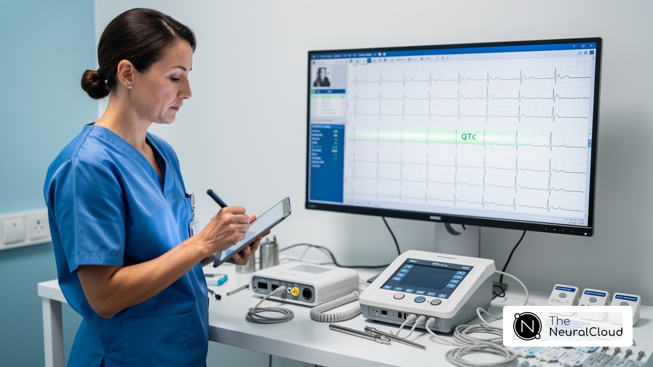
Misinterpretation of concave and convex ST elevation can lead to critical errors in ECG analysis, impacting patient care. Let’s explore the nuances of ST elevation morphology together, offering insights into their definitions, clinical implications, and the advanced tools available for precise analysis.
So, how can clinicians accurately interpret these patterns to prevent misdiagnosis and ensure the best care for their patients?
Define Concave and Convex ST Elevation
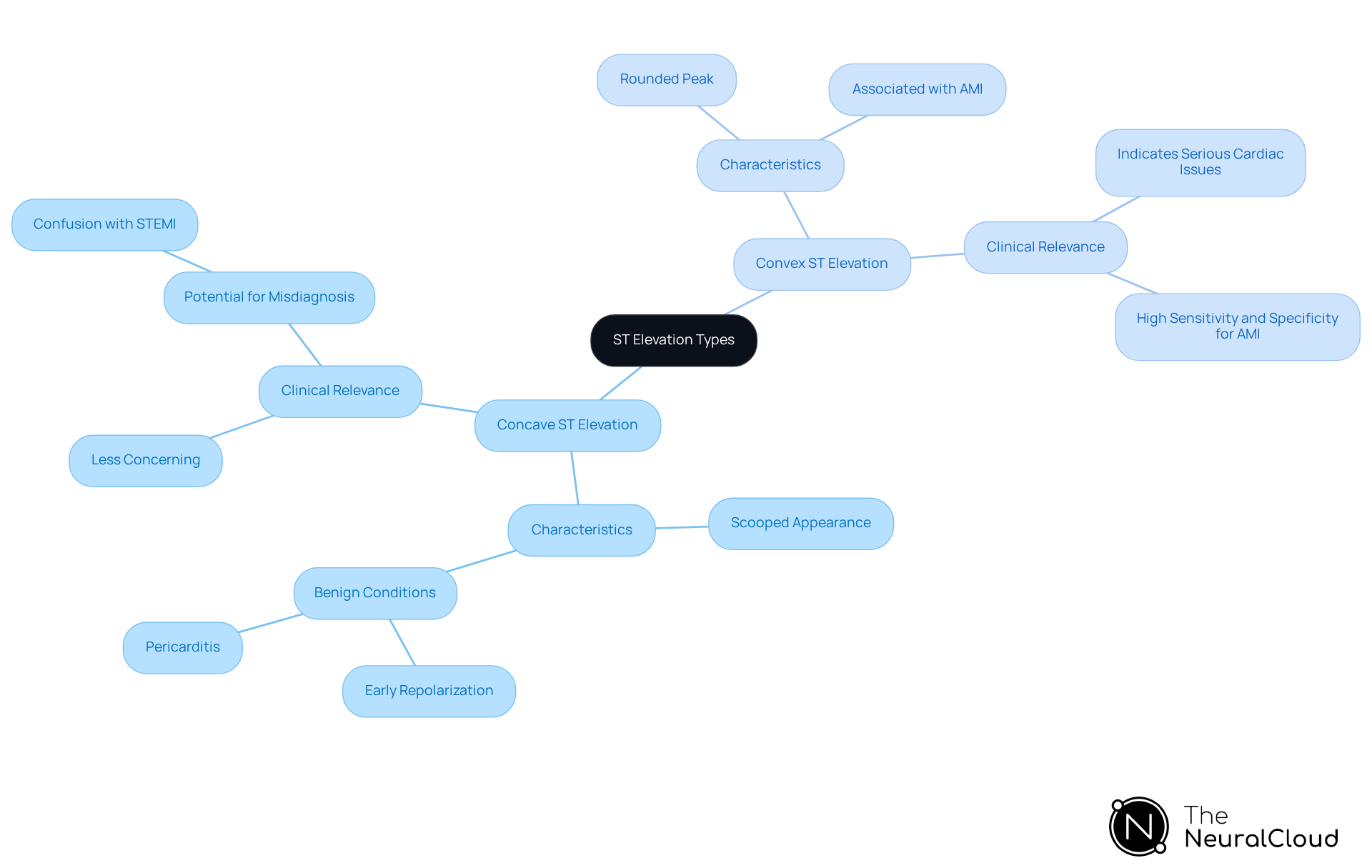
Differentiating between concave vs convex ST elevation is essential for accurate ECG interpretation, as it directly impacts clinical decision-making. The concave vs convex ST elevation is characterized by an upward-curving shape, resembling a 'scooped' appearance. This morphology is often associated with benign conditions such as early repolarization and pericarditis. In contrast, the concave vs convex ST elevation presents as a rounded peak, which is typically indicative of acute myocardial infarction (AMI) or other serious cardiac events. Understanding these definitions is crucial for accurate ECG interpretation, especially when considering concave vs convex ST elevation, as they guide clinicians in differentiating between benign and pathological conditions.
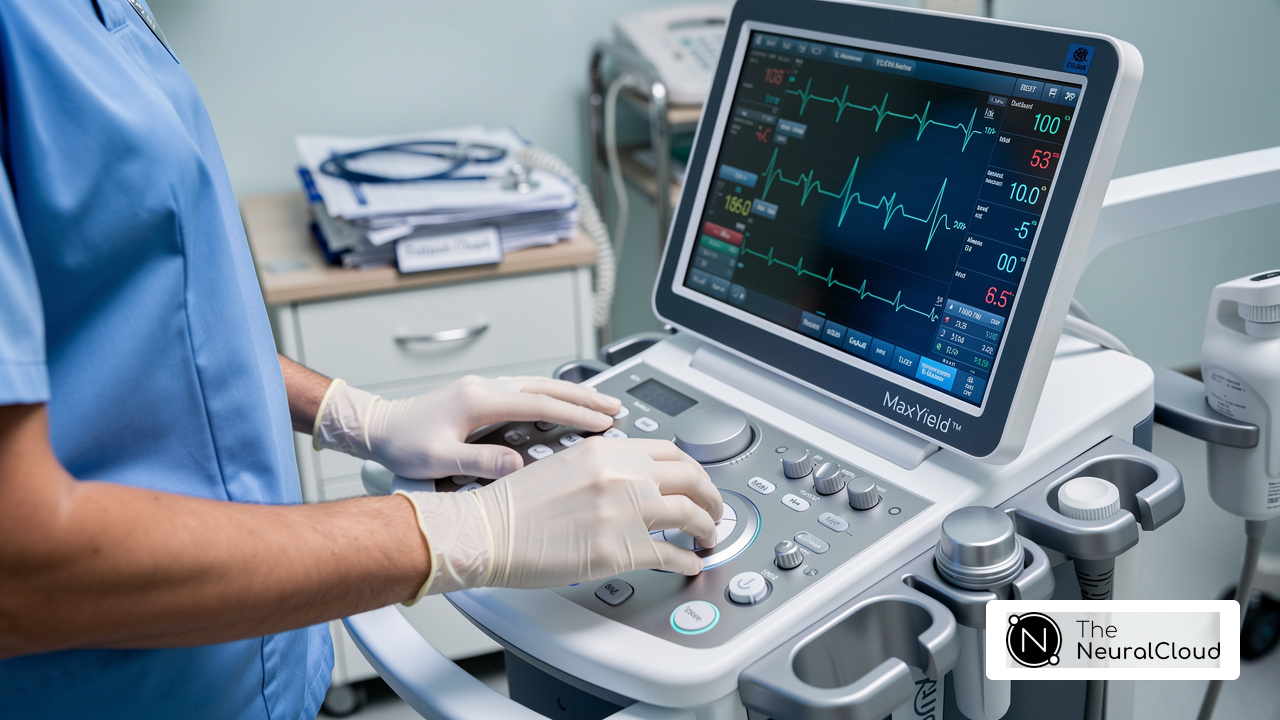
With MaxYield™, clinicians can easily map ECG signals through noise, pinpointing and labeling key features in every heartbeat. Its advanced noise filtering capabilities allow for the identification and labeling of critical data, even in recordings with high levels of noise and artifact. This guarantees that clinicians can confidently analyze ST rise configurations without the interference of baseline wander or muscle artifacts.
Key Characteristics:
- Concave ST Elevation:
- Appears 'scooped' or 'smiling'.
- Commonly seen in benign early repolarization and pericarditis.
- Appears 'frowning' or peaked.
- Often associated with acute myocardial infarction (AMI).
Clinical Relevance:
Spotting these patterns can really change the game for clinical decisions. In the context of concave vs convex ST elevation, concave protrusions are generally less concerning, while convex formations may indicate critical cardiac issues. Recent studies indicate that up to 15% of the general population may exhibit non-ischemic ST rise, complicating the interpretation of ECGs. For example, early repolarization, a harmless variant, can closely resemble ST increases observed in STEMI, highlighting the necessity for meticulous ECG analysis. Additionally, case studies highlight the importance of distinguishing between concave vs convex ST elevation morphologies to avoid unnecessary interventions and ensure appropriate treatment. With this advanced tool, clinicians can swiftly evaluate up to 200,000 heartbeats in under 5 minutes, offering prompt insights that aid assured clinical decision-making. Ultimately, with MaxYield™, clinicians can enhance their diagnostic accuracy, improving patient outcomes and reducing the risk of misdiagnosis.
Explore Clinical Significance of ST Elevation Patterns
Accurate interpretation of concave vs convex ST elevation trends is essential for diagnosing acute coronary syndromes and other cardiac conditions. With Neural Cloud Solutions' platform, examining these trends is significantly enhanced through advanced noise filtering and unique wave recognition capabilities.
Clinical Implications:
- Concave ST Elevation:
Typically benign, often associated with conditions such as early repolarization and pericarditis. Misdiagnosis can lead to unnecessary treatments, emphasizing the importance of precise analysis in ECG readings, especially in recordings with high levels of noise and artifact. - Convex ST Elevation:
Strongly linked to acute myocardial infarction (AMI), indicating potential transmural ischemia. This trend requires prompt medical action to reduce risks associated with heart injury, and can be precisely recognized even in challenging recordings due to advanced technology.
Diagnostic Considerations:
- Differential Diagnosis:
Concave patterns may lead to unnecessary interventions if misidentified, highlighting the need for accurate assessment. This system can uncover hidden parts of long Holter and patch monitor recordings, making sure no important data slips through the cracks. Conversely, convex shapes serve as critical indicators for STEMI, guiding urgent treatment decisions that can significantly impact patient outcomes. The system enhances the reliability of these interpretations by evolving its algorithms with each use.
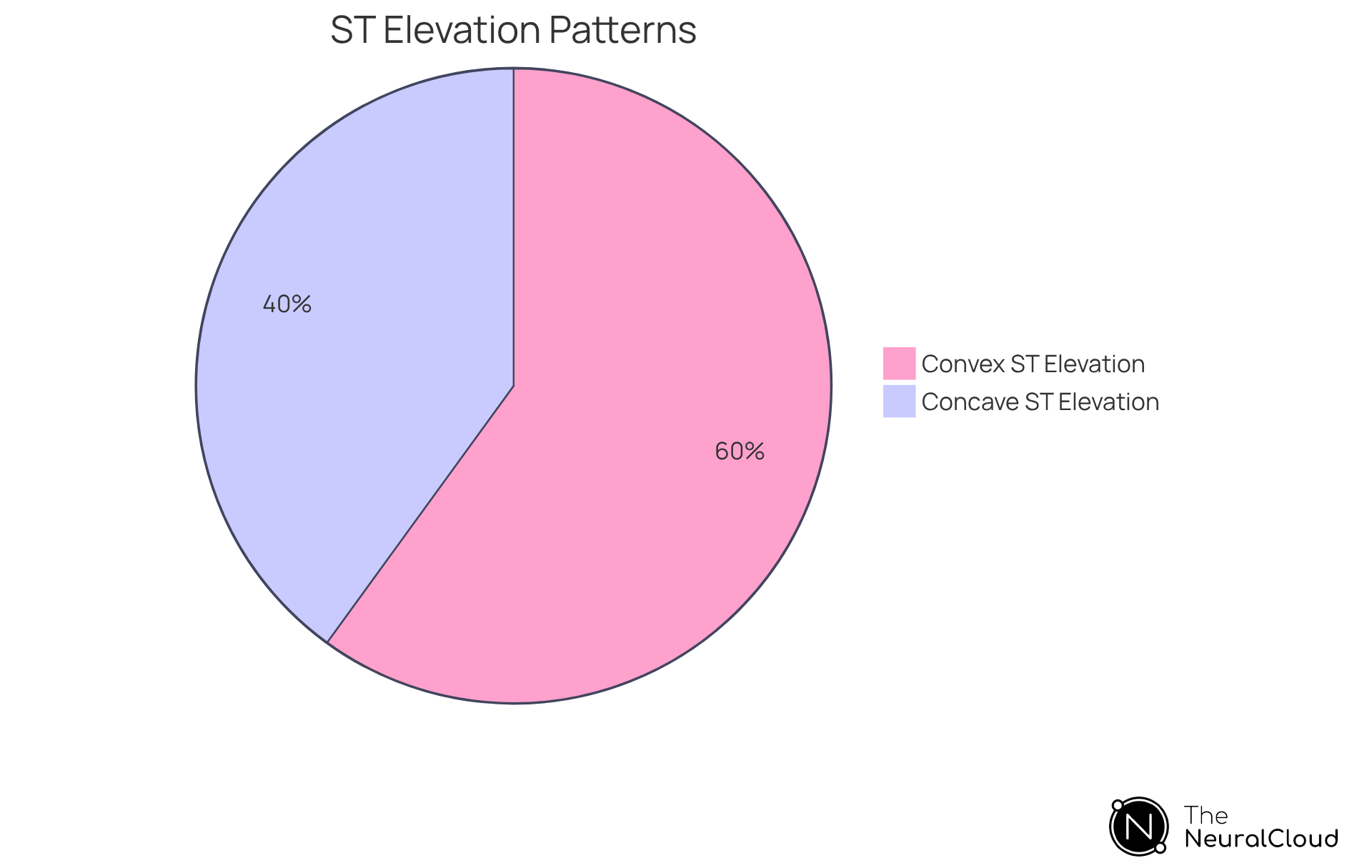
Understanding the clinical significance of concave vs convex ST elevation patterns is vital for effective risk stratification and management of patients presenting with chest pain or other cardiac symptoms. Leveraging advanced technology in ECG analysis can transform patient care by ensuring timely and accurate diagnoses.
Measure and Interpret ST Segment Morphology
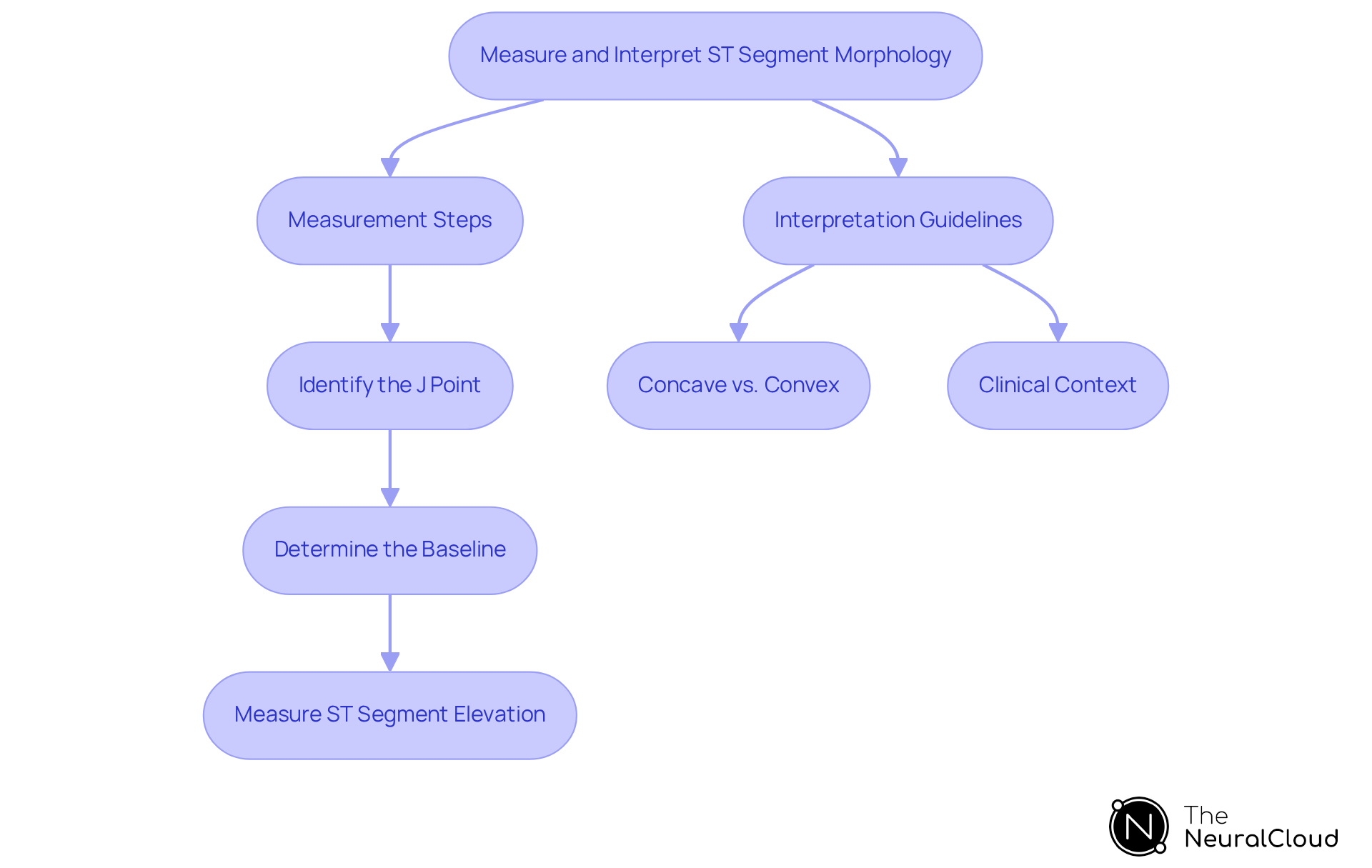
Accurate ECG analysis hinges on the precise measurement and interpretation of ST segment morphology, especially for conditions like STEMI. With the incorporation of the new system, healthcare professionals can utilize advanced ECG analysis features that improve automation, precision, and flexibility. Here are the key steps and best practices to follow:
Steps for Measurement:
- Identify the J Point:
The J point marks the transition from the S wave to the ST segment. Precise identification is essential for accurate measurement. - Determine the Baseline:
Establish a baseline by drawing a line between two TP segments, which represents the isoelectric line. - Measure ST Segment Elevation:
Measure the height of the ST segment at the J point compared to the baseline. Elevation is defined as greater than 0.1 mV above the baseline.
Interpretation Guidelines:
- Concave vs. Convex:
Assess the shape of the ST segment. Concave shapes are generally less concerning, while convex shapes may indicate serious conditions such as acute coronary occlusion. - Clinical Context:
When interpreting ST segment changes, keep clinical symptoms and other ECG findings in mind to help prevent misdiagnosis. For instance, new ST elevation with concerning symptoms should be treated as potentially serious until proven otherwise.
Practical Tips:
- Consider using software tools like MaxYield™ and Insight360 for automated measurements and customizable visualizations that can boost your accuracy and minimize human error. MaxYield™ maps ECG signals through noise, isolating and labeling key features in every heartbeat, allowing for rapid analysis of 200,000 heartbeats in less than 5 minutes.
- Regularly practice measuring ST segments on various ECGs to improve proficiency and confidence in interpretation.
- Stay informed about the latest guidelines and technological advancements in ECG analysis to ensure best practices are followed, including regulatory compliance details such as FDA Class II SaMD clearance-pending status.
Case Studies:
- Understanding ST Elevation and Its Clinical Significance:
Timely diagnosis of ST elevation is critical as it signals potential blockage in a coronary artery, which can compromise blood flow to the heart. The American College of Cardiology emphasizes performing an ECG at the first point of medical contact to enable swift diagnosis and treatment. - Differentiating ST Elevation Causes:
ST-segment elevation can arise from various conditions, including acute coronary occlusion, early repolarization, and pericarditis. Clinicians must consider the clinical context and associated symptoms when evaluating ST changes to avoid misdiagnosis. Embracing advanced tools like MaxYield™ not only streamlines the analysis process but also empowers healthcare professionals to make timely and informed decisions.
Utilize Advanced Tools for Enhanced ECG Analysis
Healthcare professionals often struggle with the accuracy and speed of ECG interpretation due to manual processes and the potential for human error. Incorporating advanced tools into ECG analysis significantly enhances the accuracy and efficiency of interpreting ST segment morphology.
Advanced Tools and Technologies:
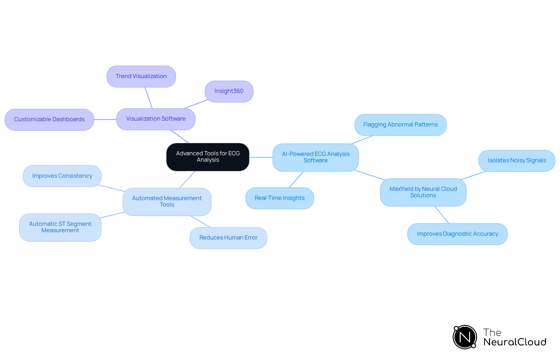
- AI-Powered ECG Analysis Software:
These tools leverage machine learning algorithms to analyze ECG data, providing real-time insights and flagging abnormal patterns. For instance, MaxYield by Neural Cloud Solutions enhances ECG data processing by isolating and cleaning noisy signals, which improves diagnostic accuracy. - Automated Measurement Tools:
Devices that automatically measure ST segment elevation help reduce human error and improve consistency in readings. - Visualization Software:
Tools like Insight360 facilitate customizable dashboards and reports, making it easier to visualize trends and interpret data effectively.
Benefits of Using Advanced Tools:
- Increased Diagnostic Accuracy:
AI tools can spot subtle changes in ST segment morphology that might slip past the human eye. For example, studies have shown that AI models can achieve a sensitivity of 92% in detecting ST-Elevation Myocardial Infarction (STEMI), compared to 71% for standard triage methods. - Time Efficiency:
These automated tools make the analysis process smoother, letting healthcare professionals concentrate on patient care instead of getting bogged down in manual calculations. The integration of AI-driven ECG analysis has been shown to reduce false positive activation rates significantly, enhancing the overall workflow in clinical settings.
Integrating advanced technologies into ECG analysis not only enhances diagnostic capabilities but also empowers healthcare professionals to provide better patient care.
Conclusion
Misunderstanding ST elevation types can lead to critical errors in ECG interpretation. Understanding the differences between concave and convex ST elevation is crucial for accurate ECG interpretation and effective clinical decision-making. By recognizing these distinct morphologies, healthcare professionals can better differentiate between benign conditions and serious cardiac events, ultimately leading to improved patient outcomes.
The article highlights the key characteristics of concave and convex ST elevation, emphasizing their clinical relevance. Concave ST elevation is often associated with benign conditions like early repolarization, while convex ST elevation is a critical indicator of acute myocardial infarction. The importance of precise measurement and interpretation of ST segment morphology is underscored, along with the role of advanced tools like MaxYield™ and Insight360 in enhancing diagnostic accuracy and efficiency.
Using advanced technologies in ECG analysis makes interpretation easier and helps clinicians make quick, informed decisions. This commitment to precision not only enhances diagnostic capabilities but also directly impacts patient care quality. With cardiology constantly changing, adopting these innovations is key to providing the best patient care. By prioritizing accurate ECG interpretation and leveraging advanced tools, healthcare professionals can significantly enhance their diagnostic capabilities and ultimately improve patient outcomes in the Canadian context.
Frequently Asked Questions
What is the difference between concave and convex ST elevation in ECG interpretation?
Concave ST elevation appears 'scooped' or 'smiling' and is often associated with benign conditions like early repolarization and pericarditis. In contrast, convex ST elevation appears 'frowning' or peaked and is typically indicative of acute myocardial infarction (AMI) or other serious cardiac events.
Why is it important to differentiate between concave and convex ST elevation?
Differentiating between concave and convex ST elevation is crucial for accurate ECG interpretation, as it directly impacts clinical decision-making. Concave patterns are generally less concerning, while convex formations may indicate critical cardiac issues.
What are the key characteristics of concave ST elevation?
Concave ST elevation appears 'scooped' or 'smiling' and is commonly seen in benign conditions such as early repolarization and pericarditis.
What are the key characteristics of convex ST elevation?
Convex ST elevation appears 'frowning' or peaked and is often associated with acute myocardial infarction (AMI).
How does MaxYield™ assist clinicians in analyzing ST elevation?
MaxYield™ helps clinicians map ECG signals through noise, allowing for the identification and labeling of critical data even in recordings with high levels of noise and artifact. This enhances the analysis of ST rise configurations without interference from baseline wander or muscle artifacts.
What is the clinical relevance of recognizing concave vs convex ST elevation?
Recognizing these patterns can significantly influence clinical decisions. For instance, up to 15% of the general population may exhibit non-ischemic ST rise, complicating ECG interpretation. Accurate identification helps avoid unnecessary interventions and ensures appropriate treatment.
How quickly can clinicians evaluate heartbeats using MaxYield™?
With MaxYield™, clinicians can swiftly evaluate up to 200,000 heartbeats in under 5 minutes, providing prompt insights that aid in clinical decision-making.
What is the impact of accurate ECG interpretation on patient outcomes?
Enhanced diagnostic accuracy through tools like MaxYield™ improves patient outcomes and reduces the risk of misdiagnosis, ensuring that patients receive appropriate care based on their ECG results.
List of Sources
- Define Concave and Convex ST Elevation
- The ST Segment (https://litfl.com/st-segment-ecg-library)
- STEMI Mimics: ECG Impostors of Heart Attack (https://powerfulmedical.com/blog/stemi-mimics)
- Checking your browser - reCAPTCHA (https://pmc.ncbi.nlm.nih.gov/articles/PMC4209433)
- Convex and Concave Morphology – RCEMLearning EMFP (https://rcemlearning.org/modules/st-elevation-without-infarction/lessons/detailed-ecg-interpretation/topic/convex-and-concave-morphology)
- Explore Clinical Significance of ST Elevation Patterns
- What Is a STEMI? (https://my.clevelandclinic.org/health/diseases/22068-stemi-heart-attack)
- STEMI ECG Criteria: ST-Elevation Thresholds (https://powerfulmedical.com/blog/stemi-ecg-criteria)
- Acute ST-Segment Elevation Myocardial Infarction (STEMI) - StatPearls - NCBI Bookshelf (https://ncbi.nlm.nih.gov/books/NBK532281)
- Cover Story | Acute Coronary Syndromes: New Perspectives, New Data - American College of Cardiology (https://acc.org/latest-in-cardiology/articles/2024/06/01/01/42/cover-story-acute-coronary-syndromes-new-perspectives-new-data)
- Measure and Interpret ST Segment Morphology
- STEMI ECG Criteria: ST-Elevation Thresholds (https://powerfulmedical.com/blog/stemi-ecg-criteria)
- pmc.ncbi.nlm.nih.gov (https://pmc.ncbi.nlm.nih.gov/articles/PMC11491539)
- ST-Segment Elevation: Definition, ECG Criteria, and Clinical Significance (https://aclscertification.org/what-is-ecg-st-segment-elevation)
- theneuralcloud.com (https://theneuralcloud.com/post/4-best-practices-for-st-elevation-v2-analysis-in-healthcare)
- Utilize Advanced Tools for Enhanced ECG Analysis
- Checking your browser - reCAPTCHA (https://pubmed.ncbi.nlm.nih.gov/41158088)
- AI ECG Interpretation Rivals Cardiologists (https://powerfulmedical.com/blog/how-is-ai-transforming-ecg-interpretation)
- AI-Based ECG Analysis Significantly Improves STEMI Detection, Reduces False Activations - American College of Cardiology (https://acc.org/latest-in-cardiology/articles/2025/10/24/16/56/tues-554pm-ai-tct-2025)