Introduction
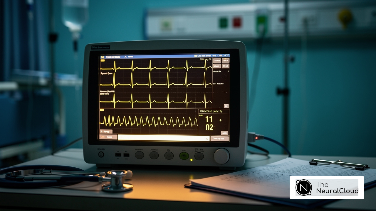
In emergency medicine, recognizing hyperacute T waves is essential for timely intervention in heart attack cases. These distinctive T waves, characterized by their pronounced amplitude and unique morphology, serve as early indicators of acute myocardial ischemia, often appearing before more obvious signs on an ECG. As the stakes rise, so does the need for healthcare professionals to master the criteria for recognizing these critical patterns. Clinicians must remain vigilant to ensure they do not miss these critical signals in high-pressure situations.
Define Hyperacute T Waves and Their Clinical Importance
In the fast-paced world of emergency medicine, recognizing the hyperacute T waves criteria is vital for effective heart attack diagnosis. HATPs are characterized by their high amplitude and wide, peaked shape, usually appearing in the initial stages of ST-elevation heart attack (STEMI). These T waves are critical indicators of acute myocardial ischemia and can often be identified according to the hyperacute T waves criteria, surfacing before other ECG changes, such as ST-segment elevation. Recognizing them quickly can lead to timely interventions that greatly improve patient outcomes. In emergency settings, rapid diagnosis is often challenging, making the identification of HATWs critical for effective treatment.
The Neural Cloud Solutions platform enhances the identification of HATWs by automating ECG analysis and filtering out noise, allowing for clearer visualization of these critical features. The system maps ECG signals through noise, isolating and labeling key features in every heartbeat, which is essential for recognizing HATWs amidst potential artifacts. With the capability to analyze 200,000 heartbeats in less than 5 minutes, MaxYield™ provides detailed insights that are crucial, especially in recordings with high levels of noise, where traditional ECG interpretation may struggle.
Recent studies have validated the HATW score, demonstrating high specificity of 98.4% and reasonable sensitivity of 20.7% for identifying obstructive heart ischemia (OMI) in patients without traditional STEMI criteria. This score emphasizes the importance of T-wave magnitude and symmetry over simple amplitude measurements, providing a more reliable diagnostic tool. For instance, the derivation group included 2,079 ECGs from 1,261 patients, with a notable prevalence of OMI at 21% and STEMI at 7.4%, underscoring the clinical utility of the HATW score in acute coronary syndrome (ACS) scenarios.
Cardiologists and other specialists emphasize the importance of hyperacute T waves criteria for diagnosing heart attacks. They note that recognizing these patterns helps guide management strategies, which can ultimately improve patient outcomes. Both the American College of Cardiology and the European Society of Cardiology recommend treating HATWs like STEMI, which means urgent reperfusion therapy is necessary. This endorsement reflects the growing consensus on the critical role of HATWs in acute myocardial ischemia diagnosis, reinforcing the need for their integration into standard ECG interpretation practices. Furthermore, it is crucial to acknowledge that one in two heart attacks are misdiagnosed at the first point of contact, emphasizing the urgency and importance of accurate HATW identification.

Outline Criteria for Identifying Hyperacute T Waves in ECGs
Accurate identification of hyperacute T waves criteria is crucial for timely diagnosis in acute coronary conditions. To assist clinicians, MaxYield™ provides specific criteria to consider:
- Amplitude: Hyperacute T waves typically exhibit a height greater than 10 mm in men and 8 mm in women, relative to the QRS complex. MaxYield™ enhances this identification by filtering out noise, ensuring that true amplitudes are accurately measured.
- Morphology: These T patterns are characterized by a broad base and symmetrical peak, often appearing more pronounced in the leads corresponding to the affected coronary artery. With MaxYield™, clinicians can clearly visualize these morphological characteristics, even in challenging recordings.
- Lead Distribution: The hyperacute T waves criteria suggest that hyperacute T deflections are usually observed in adjacent leads, particularly in the anterior leads (V2-V4) during acute heart ischemia. The system helps isolate these leads, allowing for a more focused ECG analysis.
- Temporal Changes: Sharp T patterns can develop quickly, often progressing to ST-segment elevation as myocardial damage advances. Clinicians should compare current ECGs with previous recordings to identify any new changes. With MaxYield™, you can easily compare historical data with current readings for a clearer picture.
Identifying sharp T deflections is crucial. They can lead to a diagnosis that is three hours faster in cases of acute coronary blockage. The HATW score has achieved 98.4% specificity for detecting acute coronary occlusion in patients lacking STEMI criteria, highlighting the diagnostic precision of these signals. Additionally, very sharp T patterns have twice the sensitivity in identifying occlusive myocardial infarction, making them a key aspect of ECG analysis.
MaxYield™ improves this process by offering advanced noise filtering and signal recognition features, enabling clinicians to isolate and label extremely acute T signals even in recordings with high levels of noise and artifact. This automated analysis not only enhances diagnostic yield but also supports confident clinical decision-making. Clinicians should also be aware that very early T patterns can be confused with T-shape alterations linked to high potassium levels, which present with narrow, peaked T-shapes. This emphasizes the significance of thorough assessment.

Apply Hyperacute T Waves Criteria in ECG Interpretation
Accurate interpretation of ECGs is essential for timely diagnosis and treatment of cardiac conditions, yet many healthcare professionals face challenges in this area. When interpreting ECGs for hyperacute T waves, adhere to the following steps:
- Obtain a Baseline ECG: Always compare the current ECG with previous recordings to identify any new changes in T shape morphology, which is crucial for accurate diagnosis.
- Identify Lead Placement: Focus on leads V2 to V4, where hyperacute T patterns are most pronounced, particularly in cases of anterior wall ischemia. MaxYield™ improves this process by offering accurate lead placement direction, ensuring optimal detection of these essential signals.
- Assess Amplitude and Morphology: Measure the height of the T signals and evaluate their shape. Look for broad, peaked T signals that exceed established amplitude thresholds, indicating potential occlusion. The advanced noise filtering capabilities of this system help isolate these features even in challenging recordings, improving diagnostic accuracy.
- Evaluate Clinical Context: Consider the patient's symptoms and clinical history. The occurrence of pronounced T deflections in a patient with chest discomfort should raise concerns for acute myocardial infarction, as these deflections are frequently linked to serious cardiac incidents. This system supports this evaluation by providing comprehensive analytics that correlate T changes with clinical data.
- Document Findings: Clearly record the presence of sharp T deflections in the ECG report, noting their characteristics and any associated observations, such as ST-segment changes. Keeping thorough documentation helps ensure effective patient management and informed treatment decisions, and MaxYield™ facilitates efficient reporting through automated labeling features.
In clinical settings, getting lead positioning just right is key to spotting early T formations effectively. For example, a study demonstrated that among patients lacking STEMI criteria but with a positive acute T score, 84% had a responsible lesion leading to acute myocardial infarction. This underscores the importance of precise ECG interpretation in identifying critical cardiac conditions early. Cardiologists stress that the hyperacute T waves criteria are regional and appear enlarged, featuring a wide base and round peak, which makes them a crucial indicator of occlusion. By following these guidelines and utilizing MaxYield™, healthcare professionals can significantly enhance their diagnostic precision, ultimately leading to better patient care and outcomes.

Connect Hyperacute T Waves to Occlusion Myocardial Infarction Diagnosis
Hyperacute T signals serve as critical early indicators of occlusive myocardial infarction, highlighting the urgent need for accurate ECG analysis in clinical settings. These signals indicate significant myocardial ischemia, typically resulting from coronary artery occlusion. Their identification is essential, as elevated T patterns can manifest before ST-segment elevation, serving as a vital marker for early diagnosis and intervention. Recognizing these fluctuations enables prompt reperfusion therapy, which is crucial for improving patient outcomes.
Research indicates that pronounced T deflections often appear in patients with acute chest pain, emphasizing the need for healthcare providers to remain vigilant for signs of occlusive myocardial infarction. Recent findings suggest that very early T patterns may surpass the diagnostic precision of conventional STEMI standards, underscoring their importance in identifying patients at risk of heart attacks. Notably, about half of high-risk individuals with occlusive myocardial infarction do not meet traditional STEMI criteria, making the identification of rapid T deflections a critical diagnostic tool.
Integrating the MaxYield™ platform from Neural Cloud Solutions allows healthcare professionals to significantly enhance diagnostic precision. This innovative solution automates the identification and labeling of hyperacute T waves criteria, even in noisy recordings, ensuring that clinicians can make confident decisions based on accurate data. Its adaptive algorithms continuously improve diagnostic yield, making it invaluable for the timely detection of occlusive myocardial infarction. Leveraging this advanced platform can significantly optimize treatment strategies for patients experiencing acute coronary events, ultimately saving lives in acute coronary situations.

Conclusion
The subtlety of hyperacute T waves (HATWs) presents a significant challenge in ECG interpretation, yet their early detection is crucial for timely diagnosis of myocardial ischemia. Recognizing these unique T waves acts as early warning signs for acute coronary events, often appearing before other significant changes on an ECG. Spotting them quickly can really boost patient outcomes by allowing for faster medical help, highlighting the importance for healthcare professionals to master the criteria associated with HATWs.
This article emphasizes several key insights, including the precise criteria for identifying hyperacute T waves, such as:
- Amplitude
- Morphology
- Lead distribution
Advanced tools like the MaxYield™ platform enhance the accuracy of T wave detection, even amidst noisy recordings. Additionally, the validation of the HATW score reinforces its critical role in diagnosing occlusive myocardial infarction, particularly in patients who may not present with classic STEMI criteria. By adopting these guidelines and leveraging technology, clinicians can significantly enhance their diagnostic capabilities.
In emergency medicine, the ability to swiftly and accurately identify hyperacute T waves cannot be overstated. As research continues to validate their significance, it becomes imperative for healthcare providers to integrate these criteria into standard ECG interpretation practices. Emphasizing the importance of HATWs in diagnosing myocardial infarction not only improves patient care but also highlights the necessity for ongoing education and training in ECG analysis. By prioritizing the mastery of hyperacute T waves criteria, healthcare professionals can make informed decisions that ultimately save lives in critical situations.
Frequently Asked Questions
What are hyperacute T waves (HATWs) and why are they clinically important?
Hyperacute T waves are characterized by high amplitude and a wide, peaked shape, typically appearing in the early stages of ST-elevation heart attack (STEMI). They are critical indicators of acute myocardial ischemia and can be identified before other ECG changes, facilitating timely interventions that improve patient outcomes.
How does the Neural Cloud Solutions platform assist in identifying hyperacute T waves?
The Neural Cloud Solutions platform automates ECG analysis and filters out noise, enabling clearer visualization of hyperacute T waves. It can analyze 200,000 heartbeats in less than 5 minutes, providing detailed insights that are particularly valuable in noisy recordings where traditional ECG interpretation may be challenging.
What is the significance of the HATW score in diagnosing obstructive heart ischemia (OMI)?
The HATW score has been validated to have a specificity of 98.4% and a sensitivity of 20.7% for identifying OMI in patients without traditional STEMI criteria. It emphasizes T-wave magnitude and symmetry, making it a more reliable diagnostic tool for acute coronary syndrome scenarios.
What do recent studies reveal about the prevalence of obstructive heart ischemia and STEMI?
In a study involving 2,079 ECGs from 1,261 patients, the prevalence of obstructive heart ischemia was found to be 21%, while STEMI prevalence was 7.4%. This highlights the clinical utility of the HATW score in diagnosing acute coronary syndrome.
What recommendations do cardiologists and medical organizations make regarding hyperacute T waves?
Cardiologists emphasize the importance of recognizing hyperacute T waves for diagnosing heart attacks and guiding management strategies. Both the American College of Cardiology and the European Society of Cardiology recommend treating HATWs like STEMI, necessitating urgent reperfusion therapy.
Why is accurate identification of hyperacute T waves crucial in emergency medicine?
Accurate identification of hyperacute T waves is crucial because misdiagnosis occurs in one in two heart attacks at the first point of contact. Recognizing these patterns can lead to timely and effective treatment, ultimately improving patient outcomes.
List of Sources
- Define Hyperacute T Waves and Their Clinical Importance
- powerfulmedical.com (https://powerfulmedical.com/blog/hyperacute-t-waves)
- pmc.ncbi.nlm.nih.gov (https://pmc.ncbi.nlm.nih.gov/articles/PMC12791876)
- Outline Criteria for Identifying Hyperacute T Waves in ECGs
- pmc.ncbi.nlm.nih.gov (https://pmc.ncbi.nlm.nih.gov/articles/PMC12791876)
- powerfulmedical.com (https://powerfulmedical.com/blog/hyperacute-t-waves)
- annemergmed.com (https://annemergmed.com/article/S0196-0644(05)81792-5/fulltext)
- Apply Hyperacute T Waves Criteria in ECG Interpretation
- ECG Cases 21: Hyperacute T-waves and Occlusion MI (https://emergencymedicinecases.com/hyperacute-t-waves-occlusion-mi)
- powerfulmedical.com (https://powerfulmedical.com/blog/hyperacute-t-waves)
- pmc.ncbi.nlm.nih.gov (https://pmc.ncbi.nlm.nih.gov/articles/PMC12791876)
- journalfeed.org (https://journalfeed.org/article-a-day/2023/actually-hyperacute-t-waves-can-help-identify-occlusion-mi)
- Connect Hyperacute T Waves to Occlusion Myocardial Infarction Diagnosis
- journalfeed.org (https://journalfeed.org/article-a-day/2023/actually-hyperacute-t-waves-can-help-identify-occlusion-mi)
- annemergmed.com (https://annemergmed.com/article/S0196-0644(24)01250-2/fulltext)
- physiciansweekly.com (https://physiciansweekly.com/post/early-diagnosis-of-acute-myocardial-infarction-with-hyperacute-t-waves)