Introduction
Interpreting junctional rhythms can be a daunting task for healthcare professionals, especially when subtle variations can lead to misdiagnosis. These rhythms, often indicative of underlying sinoatrial node dysfunction, present unique challenges that can significantly affect patient care.
In this guide, we’ll break down junctional rhythm ECG strip analysis step by step, ensuring you can accurately recognize and interpret these vital patterns. Recognizing these pitfalls is essential for ensuring optimal patient care and improving outcomes in ECG analysis.
Understand Junctional Rhythms: Definitions and Types
Junctional patterns present unique challenges in ECG analysis, often stemming from SA node dysfunction. You might encounter these rhythms in various forms:
- Junctional Escape Rhythm: Typically occurs at a heart rate of 40-60 beats per minute (bpm). In this pattern, P waves may be absent, inverted, or appear after the QRS complex, indicating reliance on the AV node for pacing.
- Accelerated Junctional Activity: Characterized by a heart rate between 60-100 bpm, this pattern often results from increased vagal tone or other physiological changes, reflecting the heart's adaptive response to varying conditions.
- Nodal Tachycardia: Defined as a heart rate exceeding 100 bpm, nodal tachycardia can signal underlying cardiac issues and necessitates thorough evaluation to determine the cause and appropriate management.
Recent studies suggest that sinus node dysfunction (SND) significantly contributes to junctional patterns, with approximately 20% of patients experiencing patterns attributed to SND. Recognizing the patterns on a junctional rhythm ECG strip is essential for timely and accurate diagnosis, which can significantly impact patient care.
The MaxYield™ platform integrates wearable technology to streamline ECG analysis by automating labeling and data extraction. This innovation helps address challenges such as physiological variability and signal artifacts, allowing healthcare professionals to focus on critical decision-making tasks. The MaxYield™ platform not only enhances diagnostic precision but also simplifies the recognition of junctional rhythm ECG strip patterns, ultimately aiding in improved patient outcomes.

Identify Key ECG Features of Junctional Rhythms
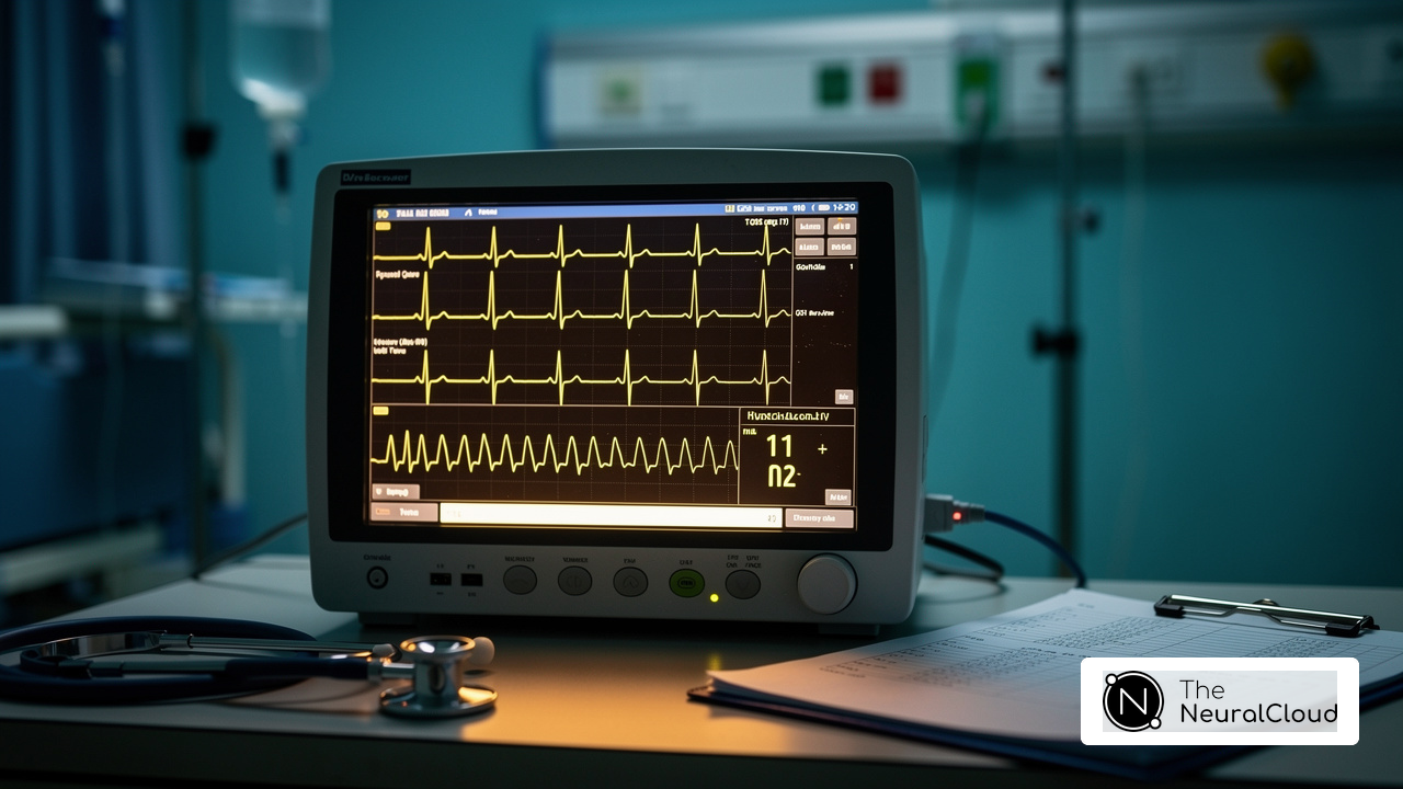
Accurate analysis of the junctional rhythm ECG strip is crucial for effective patient management in cardiology. When analyzing junctional rhythms, several key ECG features must be identified:
- P Wave Features: In connection patterns, P waves might be missing, reversed, or appear following the QRS complex. Around 40-60% of connection patterns show absent or inverted P forms, which is a hallmark characteristic that distinguishes them from sinus patterns. In premature junctional complexes (PJCs), P waves often appear inverted because of impulses traveling backward from the junction's pacemakers.
- QRS Complex: The QRS complexes in connection patterns are typically narrow, measuring less than 120 milliseconds, indicating that the impulse is conducted normally through the ventricles.
- Heart Rate: Junctional escape patterns typically present with a heart rate of 40-60 beats per minute (bpm). Accelerated junctional patterns vary from 60-100 bpm, whereas tachycardia is defined by a heart rate surpassing 100 bpm. An accelerated junction point pattern occurs when the heart rate surpasses 60 bpm, often due to enhanced automaticity or suppression of the sinoatrial node.
- Regularity: The pattern is often consistent, with steady R-R intervals, which aids in distinguishing it from other arrhythmias. During transitional patterns, the atria and ventricles depolarize at the same time, resulting in this regularity.
Recognizing the junctional rhythm ECG strip features can significantly impact patient outcomes and treatment strategies.

Follow a Step-by-Step Process for Analyzing Junctional Rhythm Strips
Analyzing a junctional rhythm ECG strip can be challenging due to noise interference and artifacts that obscure critical data. To effectively analyze these strips, adhere to the following systematic steps:
- Obtain a Clear ECG Strip: Ensure the ECG strip is of high quality, free from artifacts or noise that could obscure critical data. Utilizing the MaxYield™ platform enhances this process by filtering out noise and isolating ECG signals, even in recordings with baseline wander and muscle artifact. Proper patient preparation is essential for clarity in recordings. Ensure the patient is calm and comfortable, and that lead placement is accurate.
- Measure the Heart Rate: Count the number of R peaks in a 6-second interval and multiply by 10 to determine the heart rate. This calculation aids in classifying the rhythm as junctional escape (40-60 bpm), accelerated (60-100 bpm), or tachycardia (over 100 bpm). The MaxYield™ platform can help in accurately detecting R signals, even in noisy recordings.
- Assess P Wave Features: Examine the presence, absence, or inversion of P signals. Their position in relation to the QRS complex is vital for recognizing the particular kind of junctional rhythm ECG strip, as inverted P forms may suggest a junctional source. MaxYield™ helps clarify P wave characteristics by reducing noise interference.
- Evaluate the QRS Complex: Measure the width of the QRS complex. A standard QRS duration is less than 120 milliseconds, indicating normal conduction through the ventricles. A wide QRS may suggest underlying conduction issues. The MaxYield™ platform aids in accurately measuring QRS duration by isolating the complex from noise.
- Check Rhythm Consistency: Evaluate the consistency of the pattern by measuring the R-R intervals. Consistent intervals signify a stable connection pattern, while irregularities may indicate other cardiac conditions. MaxYield™ enhances this assessment by providing clearer interval measurements.
- Document Findings: It's crucial to document your findings accurately, as this can greatly influence clinical decisions and patient outcomes. Record your observations and interpretations meticulously, noting any abnormalities or concerns that may necessitate further investigation. The incorporation of MaxYield™ into this process highlights the significance of advanced noise filtering and signal recognition in ensuring precise ECG interpretations.
Recognizing and addressing common pitfalls in ECG interpretation can significantly enhance diagnostic accuracy and patient care. Be mindful of typical mistakes, such as incorrect lead positioning or inability to identify P characteristics, which can greatly influence analysis results.

Troubleshoot Common Issues in Junctional Rhythm Analysis
Practitioners face several challenges when analyzing junctional rhythms, which can complicate accurate interpretation and patient care. Here are some common issues that can hinder ECG analysis:
- Poor Quality ECG Strips: Practitioners often struggle with noisy or artifact-laden strips that obscure critical data. Regular calibration of the ECG machine and correct electrode placement are essential. If the strip remains unclear, re-running the ECG may be necessary to obtain a cleaner reading.
- Misinterpretation of P Currents: P currents may be absent or inverted, which can lead to confusion in diagnosis. It is essential to evaluate the connection between P waves and QRS complexes to precisely determine the kind of heart pattern. Acquaintance with distinguishing traits can assist in preventing misinterpretation, as junctional rhythm ECG strips can frequently be mistaken for other arrhythmias.
- Variable Heart Rates: Fluctuations in heart rate can complicate analysis. Employing a longer strip can assist in averaging these variations, ensuring a more precise categorization of the pattern. This approach is particularly useful in managing junctional tachycardia, which can range from 120 to 220 beats per minute.
- Inconsistent R-R Intervals: Irregularities in R-R intervals may indicate underlying issues. If the rhythm appears irregular, it is advisable to consider other arrhythmias and consult additional resources or colleagues for confirmation. This step is vital for effective cardiac monitoring and diagnosis.
- Documentation Errors: Accurate documentation of findings is critical. Double-checking notes against the ECG strip can prevent miscommunication with other healthcare providers, ensuring that all relevant information is conveyed clearly.
By being aware of these common issues and implementing the suggested solutions, healthcare professionals can significantly enhance their ECG analysis capabilities, ultimately leading to better patient outcomes. Regular updates to software and equipment maintenance are also recommended to ensure optimal system operation and accuracy in readings.

Conclusion
Mastering junctional rhythms is crucial for effective ECG analysis and optimal patient management in cardiology. By following the steps outlined in this guide, healthcare professionals can enhance their diagnostic skills and improve patient outcomes. Using advanced tools like the MaxYield™ platform makes it easier to recognize critical features in junctional rhythm ECG strips, ensuring accurate interpretations.
Key insights include:
- Various types of junctional rhythms
- Their defining characteristics
- A systematic approach for effective analysis
From identifying P wave features to evaluating heart rates and documenting findings, each step is vital for delivering precise diagnoses. Practitioners often face challenges like poor-quality ECG strips and misinterpretation of P currents, which can hinder accurate analysis. Being aware of these challenges equips practitioners with the knowledge to troubleshoot effectively and maintain high standards of care.
Ultimately, the ability to accurately analyze junctional rhythm ECG strips not only supports better clinical decision-making but also underscores the importance of continuous learning and adaptation in the field of cardiology. By embracing advanced technology and refining their analytical skills, healthcare professionals can significantly enhance patient safety and care.
Frequently Asked Questions
What are junctional rhythms in ECG analysis?
Junctional rhythms are unique patterns that arise from dysfunction of the SA node, often presenting challenges in ECG interpretation.
What is a Junctional Escape Rhythm?
A Junctional Escape Rhythm typically occurs at a heart rate of 40-60 beats per minute (bpm) and may have absent, inverted, or post-QRS complex P waves, indicating that the AV node is responsible for pacing.
What characterizes Accelerated Junctional Activity?
Accelerated Junctional Activity is characterized by a heart rate between 60-100 bpm and is often a response to increased vagal tone or other physiological changes.
What is Nodal Tachycardia?
Nodal Tachycardia is defined as a heart rate exceeding 100 bpm and may indicate underlying cardiac issues that require thorough evaluation for proper management.
How common is sinus node dysfunction (SND) in junctional rhythms?
Recent studies indicate that sinus node dysfunction (SND) significantly contributes to junctional patterns, with about 20% of patients experiencing rhythms attributed to SND.
Why is recognizing junctional rhythm patterns on ECG strips important?
Recognizing junctional rhythm patterns on ECG strips is essential for timely and accurate diagnosis, which can significantly impact patient care.
How does the MaxYield™ platform aid in ECG analysis?
The MaxYield™ platform integrates wearable technology to automate labeling and data extraction in ECG analysis, addressing challenges like physiological variability and signal artifacts, and enhancing diagnostic precision for junctional rhythms.
List of Sources
- Understand Junctional Rhythms: Definitions and Types
- researchgate.net (https://researchgate.net/publication/400205132_Junctional_Rhythm_Cardiac_Conduction_Abnormalities_and_Clinical_Implications_in_Nursing)
- journals.lww.com (https://journals.lww.com/htin/fulltext/2021/09010/a_study_on_hospitalized_patients_of_junctional.5.aspx)
- cockrell.utexas.edu (https://cockrell.utexas.edu/news/heart-rhythm-treatment-gets-breakthrough-fda-designation)
- Identify Key ECG Features of Junctional Rhythms
- theneuralcloud.com (https://theneuralcloud.com/post/master-junctional-rhythm-strip-analysis-a-step-by-step-guide)
- Junctional Rhythms (https://aclscertification.org/ecg-course-advanced-junctional-rhythms)
- Junctional Rhythm: Causes, Symptoms and Treatment (https://my.clevelandclinic.org/health/diseases/23206-junctional-rhythm)
- Follow a Step-by-Step Process for Analyzing Junctional Rhythm Strips
- emedicine.medscape.com (https://emedicine.medscape.com/article/155146-workup)
- leveluprn.com (https://leveluprn.com/blogs/ekg-interpretation/7-junctional-rhythms?srsltid=AfmBOorGYhwyAbJ5qVV76MOi5BUb60NDhNlVkzxyHr24WBBQapfCIFOa)
- theneuralcloud.com (https://theneuralcloud.com/post/master-junctional-rhythm-strip-analysis-a-step-by-step-guide)
- Electrocardiogram - StatPearls - NCBI Bookshelf (https://ncbi.nlm.nih.gov/books/NBK549803)
- practicalclinicalskills.com (https://practicalclinicalskills.com/junctional-rhythms)
- Troubleshoot Common Issues in Junctional Rhythm Analysis
- allstatesmed.com (https://allstatesmed.com/blogs/news/how-to-troubleshoot-common-ecg-machine-failures?srsltid=AfmBOopXGJ1sqA9_yMNh_ruZrrwNvusbMvG78qi_a3wGAxNt8crYbyAi)
- Junctional Rhythm: Causes, Symptoms and Treatment (https://my.clevelandclinic.org/health/diseases/23206-junctional-rhythm)
- theneuralcloud.com (https://theneuralcloud.com/post/master-junctional-rhythm-strip-analysis-a-step-by-step-guide)
- products-unlimited.com (https://products-unlimited.com/common-mistakes-in-ecg-interpretation-and-how-to-avoid-them)