Introduction
With nearly 27% of Septal Wall Myocardial Infarctions (MIs) going undiagnosed, the stakes for patient health are alarmingly high. It's essential for healthcare professionals to grasp how the interventricular septum connects with the left anterior descending artery. Timely diagnosis and intervention can drastically improve outcomes. However, the challenge for clinicians is significant: how can they effectively recognize and treat this condition to mitigate its life-threatening consequences? Recognizing this condition early can be the difference between life and death for patients.
Define Septal Wall Myocardial Infarction and Its Clinical Importance
Septal Wall Myocardial Infarction (MI) presents significant challenges due to its potential for severe complications and the need for timely intervention. This condition is characterized by the necrosis of cardiac tissue within the interventricular septum, the barrier that separates the left and right ventricles. It is primarily caused by a blockage of the left anterior descending artery (LAD), which is crucial for supplying blood to this partition area. The clinical significance of septal MI cannot be overstated, as it is associated with serious complications, including:
- Heart failure
- Arrhythmias
- The risk of sudden cardiac death
These complications create serious hurdles for healthcare providers trying to ensure the best patient outcomes.
Recent studies indicate that the incidence of definitive MI diagnosis in hospitalized individuals is approximately 272 per 100,000 people aged 30-74, highlighting the prevalence of this condition. Moreover, the worldwide occurrence of MI in individuals under 60 years is noted at 3.8%, while it increases to 9.5% for those above 60 years. This underscores the importance of acting quickly to reduce risks and improve patient outcomes. The outlook for individuals with a myocardial infarct often depends on the extent of cardiac damage and how quickly they receive treatment. Timely intervention can significantly improve outcomes by restoring blood flow and minimizing myocardial damage.
Complications resulting from ventricular MI include:
- Ventricular rupture (VSR), which can lead to further impairment of cardiac function and arrhythmias
The prognosis for VSR depends on how large the tear is and how quickly medical help is provided. Additionally, the effect of LAD occlusion on myocardial infarction outcomes in the septum is significant, as it can result in localized tissue damage that disrupts normal electrical signaling and pumping efficiency.
Understanding the implications of cardiac muscle MI is essential for healthcare professionals, as it informs both diagnostic and therapeutic strategies. Recognizing symptoms such as:
- Chest pain
- Shortness of breath
- Fatigue
is crucial for early detection and timely medical intervention. Furthermore, the high prevalence of undiagnosed MI, reported at 26.9%, emphasizes the need for vigilance in recognizing symptoms and conducting appropriate tests. Follow-up appointments after a cardiac event are also vital for monitoring recovery and ensuring ongoing care.
Overall, managing myocardial infarction in the septum, as shown by the septal wall MI ECG, requires a comprehensive approach that includes monitoring, lifestyle modifications, and possibly surgical interventions to restore optimal heart function.

Identify Key ECG Features of Septal Wall Myocardial Infarction
Accurate diagnosis and prompt intervention in myocardial infarction hinge on recognizing specific ECG characteristics. Key indicators include:
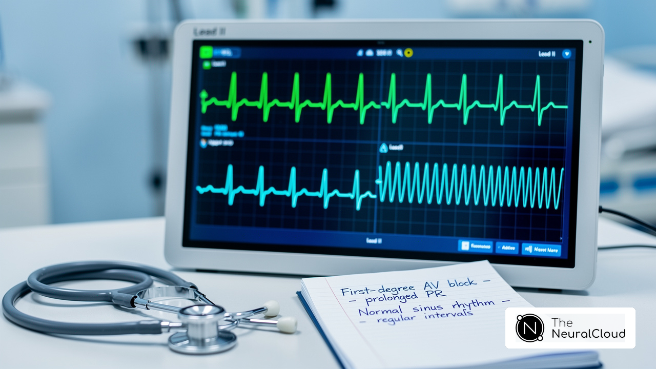
- Q Waves in Leads V1-V3: Pathological Q waves in these leads signify myocardial necrosis. In instances of myocardial infarction affecting the septal wall mi ecg, these waves are mainly observed in the precordial leads, particularly V1 and V2. Research suggests that as many as 50% of patients may show these alterations.
- ST Segment Elevation: An elevation in the ST segment in leads V1 and V2 is a hallmark of acute ischemia impacting the wall region. This finding is crucial for diagnosing an acute myocardial infarction, as it indicates underlying myocardial damage that can be assessed through the septal wall mi ecg.
- Reciprocal Changes: In certain instances, reciprocal ST segment depression may be noted in the inferior leads (II, III, aVF). This phenomenon can further support the diagnosis of myocardial infarction in the partition, highlighting the interconnected nature of cardiac electrical activity.
- Loss of R Wave Progression: A reduction in R wave amplitude in the precordial leads indicates involvement of the interventricular septum, especially in the context of anterior myocardial infarctions, which can be observed in the septal wall mi ecg. This loss of progression is a significant indicator that should not be overlooked during ECG interpretation.
Without a clear understanding of these ECG indicators, healthcare providers may struggle to make timely and accurate diagnoses of myocardial infarction. By accurately identifying these ECG features, healthcare providers can implement timely interventions that significantly improve patient outcomes. Recognizing these ECG characteristics not only aids in diagnosis but also plays a crucial role in enhancing patient care and outcomes.

Outline Diagnostic Approaches for Septal Wall Myocardial Infarction
Diagnosing septal wall myocardial infarction can be complex, requiring a thorough approach to ensure accuracy. Here are the key steps involved:
- Patient History and Symptoms: A detailed patient history is essential for accurate diagnosis. Symptoms typically include chest pain, shortness of breath, and fatigue. Understanding risk factors, such as a history of coronary artery disease, is critical, as approximately 3.1% of U.S. adults are affected by myocardial infarction, translating to about 8.8 million individuals.
- Electrocardiogram (ECG): Think of the ECG as the first step in diagnosing myocardial infarction. Specific changes in the ECG, such as ST-segment elevation or T-wave inversions, can indicate septal region MI. A 12-lead ECG should be performed immediately upon suspicion of an MI, as timely identification is crucial for effective management.
- Cardiac Biomarkers: Blood tests measuring cardiac troponins (I and T) are vital for confirming myocardial injury. Elevated levels indicate damage to the heart muscle and support the diagnosis of MI. High-sensitivity troponin tests are particularly effective, as they can detect even minor elevations in cardiac injury.
- Imaging Studies: In specific instances, echocardiography may be employed to assess motion abnormalities of the heart and evaluate the extent of myocardial damage. Advanced imaging techniques, such as cardiac MRI, can provide detailed insights into myocardial viability, aiding in treatment planning.
- Coronary Angiography: This invasive procedure visualizes the coronary arteries to identify blockages. It is especially beneficial in devising revascularization strategies, which are essential for restoring blood flow in individuals with significant coronary artery disease.
Using these diagnostic methods, healthcare providers can pinpoint myocardial infarction and start timely treatment, greatly improving patient outcomes.

Explore Treatment Strategies for Septal Wall Myocardial Infarction
Treating septal wall MI ECG presents unique challenges that require a multifaceted approach. Key components of effective treatment strategies include medications, revascularization, coronary artery bypass grafting (CABG), monitoring and supportive care, and rehabilitation.
-
Medications:
- Initial management often includes antiplatelet agents such as aspirin and clopidogrel.
- Anticoagulants are used to prevent further clot formation.
- Beta-blockers, ACE inhibitors, and statins enhance cardiac function and reduce cardiovascular risk.
- Dual antiplatelet therapy (DAPT) is essential for individuals with heart attacks, stents, or CABG, greatly lowering the risk of subsequent cardiovascular events.
-
Revascularization:
- Percutaneous coronary intervention (PCI) is the preferred approach for restoring blood flow in acute MI cases.
- This procedure involves stent insertion to clear obstructed arteries, especially in the left anterior descending (LAD) artery, which supplies blood to the region between the heart chambers.
Recent studies indicate that the primary PCI success rate in individuals with septal wall MI ECG is approximately 67.9%, compared to 92.3% in control groups, highlighting the importance of timely intervention.
-
Coronary Artery Bypass Grafting (CABG):
- In situations where PCI is not possible or for individuals with multi-vessel disease, CABG may be recommended to bypass blocked arteries and improve blood circulation to the organ.
-
Monitoring and Supportive Care:
- Continuous monitoring in a hospital setting is crucial for managing potential complications such as arrhythmias or heart failure.
- Supportive care, including oxygen therapy and fluid management, may also be necessary to stabilize the individual.
- In-hospital mortality rates for individuals with prior CABG undergoing PCI have been reported at 11.5%, compared to 2.5% in those without, underscoring the need for vigilant monitoring.
-
Rehabilitation:
- Following discharge, cardiac rehabilitation programs play a vital role in recovery.
- These programs focus on lifestyle modifications, exercise, and education to prevent future cardiovascular events.
Ultimately, a comprehensive treatment strategy can significantly improve recovery and long-term health for patients diagnosed with septal wall MI ECG. Expert insights, such as those from Dr. Alan Rabinowitz, emphasize the importance of tailored antiplatelet therapy in improving patient outcomes.

Conclusion
Timely intervention in Septal Wall Myocardial Infarction (MI) is crucial, as delays can lead to severe complications. It's vital for healthcare professionals to grasp the details of this condition, from its definition to diagnosis and treatment strategies. The insights provided throughout the article highlight the importance of recognizing key ECG features, understanding diagnostic approaches, and implementing effective treatment strategies to manage septal wall MI effectively.
Key arguments presented include the identification of specific ECG indicators such as:
- Q waves
- ST segment elevation
- Loss of R wave progression
These are vital for accurate diagnosis. Additionally, the article emphasizes the significance of a comprehensive diagnostic approach that includes:
- Patient history
- Cardiac biomarkers
- Imaging studies
Treatment strategies, ranging from medication management to revascularization techniques, are crucial for restoring blood flow and minimizing myocardial damage. Continuous monitoring and rehabilitation post-discharge play a pivotal role in long-term recovery and prevention of future cardiovascular events.
Managing Septal Wall Myocardial Infarction involves a comprehensive approach that includes timely diagnosis, effective treatment, and continuous care. Healthcare providers must remain vigilant in recognizing symptoms and utilizing appropriate diagnostic tools to ensure prompt intervention. By enhancing education and awareness, healthcare providers can significantly improve patient outcomes and reduce risks associated with septal wall MI.
Frequently Asked Questions
What is Septal Wall Myocardial Infarction (MI)?
Septal Wall Myocardial Infarction is a condition characterized by the necrosis of cardiac tissue within the interventricular septum, primarily caused by a blockage of the left anterior descending artery (LAD).
Why is Septal Wall MI clinically significant?
Septal Wall MI is clinically significant due to its association with severe complications such as heart failure, arrhythmias, and the risk of sudden cardiac death, which pose challenges for healthcare providers in ensuring optimal patient outcomes.
What is the incidence of Septal Wall MI in hospitalized individuals?
The incidence of definitive MI diagnosis in hospitalized individuals is approximately 272 per 100,000 people aged 30-74.
How does age affect the occurrence of myocardial infarction?
The worldwide occurrence of myocardial infarction in individuals under 60 years is 3.8%, while it increases to 9.5% for those above 60 years.
What are the potential complications of Septal Wall MI?
Complications can include ventricular rupture (VSR), which can impair cardiac function and lead to arrhythmias. The prognosis for VSR depends on the size of the tear and the speed of medical intervention.
What symptoms are associated with Septal Wall MI?
Symptoms include chest pain, shortness of breath, and fatigue, which are crucial for early detection and timely medical intervention.
What is the prevalence of undiagnosed myocardial infarction?
The prevalence of undiagnosed myocardial infarction is reported at 26.9%, highlighting the need for vigilance in recognizing symptoms and conducting appropriate tests.
What is the importance of follow-up appointments after a cardiac event?
Follow-up appointments are vital for monitoring recovery and ensuring ongoing care after a cardiac event.
How can the management of Septal Wall MI be approached?
Management requires a comprehensive approach that includes monitoring, lifestyle modifications, and possibly surgical interventions to restore optimal heart function.
List of Sources
- Define Septal Wall Myocardial Infarction and Its Clinical Importance
- The global prevalence of myocardial infarction: a systematic review and meta-analysis - PMC (https://pmc.ncbi.nlm.nih.gov/articles/PMC10122825)
- What Is a Septal Infarct? (https://my.clevelandclinic.org/health/diseases/septal-infarct)
- Septal infarct: Causes, symptoms, and diagnosis (https://medicalnewstoday.com/articles/septal-infarct)
- Identify Key ECG Features of Septal Wall Myocardial Infarction
- What Is a Septal Infarct? (https://my.clevelandclinic.org/health/diseases/septal-infarct)
- Acute Myocardial Infarction - StatPearls - NCBI Bookshelf (https://ncbi.nlm.nih.gov/books/NBK459269)
- Septal infarct: Causes, symptoms, and diagnosis (https://medicalnewstoday.com/articles/septal-infarct)
- ECG Learning Center - An introduction to clinical electrocardiography (https://ecg.utah.edu/lesson/9)
- theneuralcloud.com (https://theneuralcloud.com/post/understanding-septal-infarct-ecg-findings-for-effective-diagnosis)
- Outline Diagnostic Approaches for Septal Wall Myocardial Infarction
- Acute Myocardial Infarction - StatPearls - NCBI Bookshelf (https://ncbi.nlm.nih.gov/books/NBK459269)
- ACS and Cardiac Biomarkers - American College of Cardiology (https://acc.org/clinical-topics/acute-coronary-syndromes/acs-and-cardiac-biomarkers)
- Explore Treatment Strategies for Septal Wall Myocardial Infarction
- In-hospital outcome of primary PCI for patients with acute myocardial infarction and prior coronary artery bypass grafting - PMC (https://pmc.ncbi.nlm.nih.gov/articles/PMC8024815)
- Heart Attack Treatment (https://heart.org/en/health-topics/heart-attack/treatment-of-a-heart-attack)
- Myocardial Infarction: Three drug trials to watch in 2024 (https://clinicaltrialsarena.com/features/myocardial-infarction-three-trials-watch-2024)
- National Registry of Myocardial Infarction Treatment Rates (https://uclahealth.org/departments/medicine/cardiology/research/heart-failure-champ/national-registry-myocardial-infarction-treatment-rates)
- Beta Blockers, the Standard Treatment After a Heart Attack, May Offer No Benefit for Heart Attack Patients and Women Can Have Worse Outcomes | Newswise (https://newswise.com/articles/beta-blockers-the-standard-treatment-after-a-heart-attack-may-offer-no-benefit-for-heart-attack-patients-and-women-can-have-worse-outcomes)