Introduction
Recognizing the critical role of ECG analysis in diagnosing myocardial infarction is essential for effective patient care. As heart attacks continue to pose significant health risks, mastering the stages of myocardial infarction ECG can empower healthcare professionals to make timely and accurate diagnoses. However, healthcare professionals often struggle to identify these rapid changes, which can lead to misdiagnosis. By leveraging AI and advanced technologies, healthcare professionals can enhance their diagnostic capabilities, leading to improved patient outcomes.
Define Myocardial Infarction and Its ECG Relevance
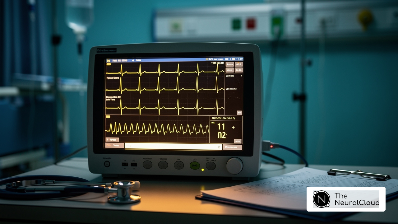
Myocardial damage, commonly known as a heart attack, poses significant challenges in timely diagnosis and treatment, making effective ECG analysis crucial. This condition occurs when blood flow to a portion of the heart is obstructed, resulting in harm or necrosis of heart muscle tissue. Frequently, blockages in one or more coronary arteries due to atherosclerosis or thrombosis cause this condition. The electrocardiogram (ECG) serves as an essential diagnostic tool for the stages of myocardial infarction ECG, revealing typical changes in the heart's electrical activity that suggest ischemia and tissue damage.
Key ECG changes associated with MI include:
- ST-segment elevation: A hallmark sign of STEMI, indicating a complete blockage of a coronary artery. Timely recognition of ST-segment elevation is critical, as immediate intervention can significantly reduce mortality rates.
- T-wave inversion: Often occurs in the stages of myocardial infarction ECG, indicating ongoing harm to the heart muscle.
- Pathological Q waves: Indicate significant heart tissue damage, typically observed in the stages of myocardial infarction ECG.
Recent studies highlight the diagnostic accuracy of ECG in identifying MI. For instance, AI-based ECG interpretation has demonstrated a sensitivity of 77% and a specificity of 99% in detecting obstructive MI, outperforming traditional diagnostic pathways, which correctly identified occlusive MI in only 42% of cases. This advancement underscores the potential of incorporating AI technologies, such as Neural Cloud Solutions' MaxYield™, into ECG analysis to improve early detection and treatment of heart attacks. MaxYield™'s advanced noise filtering and distinct wave recognition capabilities allow for the isolation of critical data even in recordings with high levels of noise and artifact, ensuring that clinicians can make confident decisions based on clear, actionable insights.
The stages of myocardial infarction ECG reveal changes that have a significant impact on patient outcomes, emphasizing the need for timely diagnosis and intervention. Ideally, treatment should occur within 90 minutes of symptom onset to improve survival rates in STEMI cases. Research indicates that delays in treatment can significantly increase the risk of mortality, highlighting the importance of rapid and accurate ECG interpretation in emergency settings. As Bryn Mumma, a UC Davis Health professor, stated, "The best outcomes happen when technology and clinicians work hand in hand, combining powerful tools with expert medical judgment." As research continues to advance, the integration of AI in ECG analysis could redefine standards of care in cardiovascular health, ultimately enhancing patient outcomes. Furthermore, experts like Dr. Alan Rabinowitz and Dr. Brett Heilbron emphasize that MaxYield™ rivals human interpretation in precision, making it an invaluable tool in the fight against heart disease. Additionally, it is important to note that MaxYield™ is currently pending FDA Class II SaMD clearance, ensuring compliance with regulatory standards.

Explore ECG Changes Across Myocardial Infarction Stages
Understanding the stages of myocardial infarction ECG changes is essential for timely and accurate diagnosis, yet these changes can be subtle and evolve over time. The ECG changes can be categorized into several critical stages:
- Hyperacute T Waves: These represent the earliest signs of an MI, emerging within minutes to hours after ischemia onset. Characterized by tall, peaked T waves in the leads corresponding to the affected area, hyperacute T waves are crucial for early diagnosis. Recent studies indicate that leads III, aVR, and V1 exhibit positive likelihood ratios of 3.8, 4.3, and 2.0, respectively. An upright T-wave amplitude exceeding the 95th percentile is indicative of vertical hyperacute T waves.
- ST-Segment Elevation: Typically occurring within the first few hours of an MI, ST-segment elevation is a hallmark of ST-Elevation Myocardial Infarction (STEMI). This elevation is most pronounced in the leads that correspond to the region of the heart impacted by the damage. Concordant ST elevation of ≥1 mm in any lead supports the diagnosis of acute MI, particularly in cases of ventricular paced rhythm (VPR).
- Q Wave Formation: As tissue damage advances, pathological Q waves may develop within hours to days. When Q waves appear, it signals that heart tissue has died, making it a key marker for diagnosing an MI. In individuals with left bundle branch block (LBBB), the Smith Modified Sgarbossa Criteria can improve the sensitivity of identifying heart attacks.
- T-Wave Inversion: Following the initial elevation, T waves may become inverted, signaling ongoing ischemia or the resolution of the acute phase of the MI. This inversion can also be observed in conditions such as Brugada syndrome, which may mimic STEMI.
- Return to Baseline: Sometimes, the ECG returns to baseline after the acute phase, but this doesn’t always mean full recovery, as some changes might still be present. Understanding the stages of myocardial infarction ECG is crucial for the precise diagnosis and management of heart attacks, particularly in differentiating between STEMI and its imitations, such as pulmonary embolism (PE) and left ventricular aneurysm (LVA). This highlights the importance of integrating advanced imaging techniques for accurate diagnosis when ECG findings are ambiguous.

Identify Diagnostic Challenges in Myocardial Infarction ECG Interpretation
Interpreting the stages of myocardial infarction ECG during myocardial infarction (MI) can be challenging, impacting the accuracy of diagnoses and patient care. Several factors contribute to this complexity:
- Variability in Presentation: Patients may exhibit various ECG changes depending on the type of MI - ST-elevation heart attack (STEMI) versus non-ST-elevation heart attack (NSTEMI) - and the location of the attack. This variability in the stages of myocardial infarction ECG can result in misdiagnosis, complicating timely treatment for patients. Approximately 40% of true STEMI patients may present with a STEMI equivalent that does not meet classic diagnostic criteria, highlighting the importance of understanding the stages of myocardial infarction ECG to avoid potential misdiagnosis.
- False Positives and Negatives: Conditions such as pericarditis or early repolarization can mimic MI on an ECG, resulting in false-positive diagnoses. Conversely, some MIs may not exhibit classic changes in the stages of myocardial infarction ECG, which can lead to false negatives. Recent studies indicate that the AI-based ECG model significantly reduced false positives for STEMI from 41.8% to 7.9%, showcasing the potential for improved diagnostic accuracy.
- Technical Factors: Poor lead placement, patient movement, or electrical interference can compromise ECG quality, leading to misinterpretation. Ensuring proper technique and equipment functionality is crucial for obtaining accurate readings. The AI ECG model has demonstrated a sensitivity of 92% and specificity of 81% in identifying the stages of myocardial infarction ECG, outperforming standard care, which had a sensitivity of only 71% and specificity of 29%.
- Subtle Changes: Early signs of MI, such as hyperacute T waves, may be subtle and easily overlooked. Clinicians must remain vigilant in recognizing these early indicators to facilitate timely intervention. The AI model can even spot occlusive MI in patients without ST elevation, showing just how crucial advanced diagnostic tools are for catching these subtle changes.
- Comorbidities: Patients with pre-existing conditions, such as bundle branch blocks or electrolyte imbalances, may present with altered ECG patterns, complicating the interpretation of MI-related changes. The variability in MI presentation necessitates a nuanced understanding of how these comorbidities can affect the stages of myocardial infarction ECG readings, emphasizing the need for advanced algorithms that can adapt to diverse patient profiles.
By leveraging advanced diagnostic tools, healthcare professionals can significantly enhance the accuracy of MI diagnoses, ultimately leading to better patient outcomes.

Utilize Advanced Technologies for Enhanced ECG Analysis
Traditional methods of ECG analysis face significant challenges, particularly in accurately detecting the stages of myocardial infarction ECG. Advanced technologies are now revolutionizing ECG analysis and interpretation, providing healthcare professionals with powerful tools to enhance diagnostic accuracy and improve patient outcomes.
- Artificial Intelligence: AI algorithms can process extensive ECG datasets, identifying patterns and abnormalities that may elude human interpreters. These systems can send real-time alerts for critical changes, which helps healthcare teams respond faster in emergencies.
- Machine Learning: Machine learning models are trained on large datasets to detect subtle variations in ECG waveforms, thereby improving MI detection accuracy. These models continuously adapt and refine their capabilities as they learn from new data, ensuring ongoing enhancement in diagnostic precision.
- Automated Interpretation Tools: Software solutions that integrate seamlessly with existing ECG machines offer automated interpretations, alleviating the workload on healthcare professionals and facilitating quicker decision-making processes.
- Telemedicine Integration: Remote monitoring and telemedicine platforms empower healthcare providers to analyze ECGs in real-time, enabling timely interventions and broadening access to care for patients, particularly in underserved areas.
- Data Visualization: Advanced visualization tools, such as Neural Cloud Solutions' Insight360, provide clinicians with customizable formats to view ECG data. This capability simplifies the identification of trends and anomalies indicative of the stages of myocardial infarction ECG, enhancing diagnostic accuracy and efficiency.
Recent studies underscore the efficacy of AI in this domain, with AI-based ECG interpretation achieving an accuracy rate of 84% in identifying obstructive MI, significantly outperforming traditional methods. The adoption of these advanced technologies not only enhances diagnostic accuracy but also transforms patient care in critical situations.

Conclusion
Accurate ECG analysis is crucial for diagnosing myocardial infarction and ensuring timely patient intervention. The insights presented emphasize the critical role of ECG in identifying the various changes that occur during an MI, which can significantly influence patient outcomes. By mastering these ECG interpretations, healthcare professionals can enhance their diagnostic capabilities and ensure that patients receive the necessary treatment as swiftly as possible.
Key ECG changes associated with myocardial infarction include:
- Hyperacute T waves
- ST-segment elevation
- Pathological Q waves
Each of these serves as a pivotal indicator of heart damage. The integration of advanced technologies, such as AI-driven ECG analysis with Neural Cloud Solutions' MaxYield™, further enhances the accuracy of these interpretations, reducing the likelihood of misdiagnosis and improving the overall patient care experience. Additionally, recognizing the challenges in ECG interpretation, from variability in presentation to technical factors, underscores the need for continuous education and the adoption of innovative diagnostic tools.
In conclusion, staying ahead of technological advancements in ECG interpretation is not just beneficial; it’s essential for improving patient survival rates in critical situations. By prioritizing accurate ECG interpretation and leveraging state-of-the-art tools, clinicians can significantly improve their response to heart attacks, ultimately saving lives and enhancing patient outcomes. The commitment to staying informed and adapting to these advancements is crucial in the ongoing fight against heart disease, ensuring that every patient receives the best possible care during critical moments.
Frequently Asked Questions
What is myocardial infarction?
Myocardial infarction, commonly known as a heart attack, occurs when blood flow to a part of the heart is obstructed, leading to damage or necrosis of heart muscle tissue.
What causes myocardial infarction?
Myocardial infarction is frequently caused by blockages in one or more coronary arteries due to atherosclerosis or thrombosis.
How is ECG relevant to diagnosing myocardial infarction?
The electrocardiogram (ECG) is an essential diagnostic tool that reveals changes in the heart's electrical activity, indicating ischemia and tissue damage associated with myocardial infarction.
What are the key ECG changes associated with myocardial infarction?
Key ECG changes include:
- ST-segment elevation, indicating complete blockage of a coronary artery.
- T-wave inversion, suggesting ongoing harm to the heart muscle.
- Pathological Q waves, indicating significant heart tissue damage.
How accurate is ECG in detecting myocardial infarction?
Recent studies show that AI-based ECG interpretation has a sensitivity of 77% and a specificity of 99% in detecting obstructive myocardial infarction, significantly outperforming traditional diagnostic methods.
What is MaxYield and how does it improve ECG analysis?
MaxYield, developed by Neural Cloud Solutions Inc., enhances ECG analysis through advanced noise filtering and distinct wave recognition capabilities, allowing for the isolation of critical data even in noisy recordings.
Why is timely diagnosis and treatment of myocardial infarction important?
Timely diagnosis and treatment, ideally within 90 minutes of symptom onset, can significantly improve survival rates in STEMI cases. Delays in treatment can increase the risk of mortality.
What does current research suggest about the integration of AI in ECG analysis?
Research indicates that integrating AI technologies like MaxYield could redefine standards of care in cardiovascular health and enhance patient outcomes by improving early detection and treatment of heart attacks.
Is MaxYield pending any regulatory approvals?
Yes, MaxYield is currently pending FDA Class II SaMD clearance, ensuring compliance with regulatory standards.
List of Sources
- Define Myocardial Infarction and Its ECG Relevance
- Heart Disease Facts (https://cdc.gov/heart-disease/data-research/facts-stats)
- AI-based ECG interpretation outperforms standard diagnosis of occlusive myocardial infarction (https://news-medical.net/news/20260323/AI-based-ECG-interpretation-outperforms-standard-diagnosis-of-occlusive-myocardial-infarction.aspx)
- New study finds AI model improves heart attack detection (https://health.ucdavis.edu/news/headlines/new-study-finds-ai-model-improves-heart-attack-detection/2025/11)
- Artificial intelligence may speed heart attack diagnosis and treatment (https://newsroom.heart.org/news/artificial-intelligence-may-speed-heart-attack-diagnosis-and-treatment)
- Explore ECG Changes Across Myocardial Infarction Stages
- physiciansweekly.com (https://physiciansweekly.com/post/early-diagnosis-of-acute-myocardial-infarction-with-hyperacute-t-waves)
- STEMI Mimics: Spot Subtle Impostors of Myocardial Infarction (https://powerfulmedical.com/blog/stemi-mimics)
- Acute ST-Segment Elevation Myocardial Infarction (STEMI) - StatPearls - NCBI Bookshelf (https://ncbi.nlm.nih.gov/books/NBK532281)
- sciencedirect.com (https://sciencedirect.com/science/article/pii/S0196064424012502)
- AI-based ECG interpretation outperforms standard diagnosis of occlusive myocardial infarction (https://news-medical.net/news/20260323/AI-based-ECG-interpretation-outperforms-standard-diagnosis-of-occlusive-myocardial-infarction.aspx)
- Identify Diagnostic Challenges in Myocardial Infarction ECG Interpretation
- healio.com (https://healio.com/news/cardiology/20251030/aibased-ecg-model-linked-to-reduction-in-false-positives-for-heart-attack)
- AI-based ECG interpretation outperforms standard diagnosis of occlusive myocardial infarction (https://news-medical.net/news/20260323/AI-based-ECG-interpretation-outperforms-standard-diagnosis-of-occlusive-myocardial-infarction.aspx)
- AI outperforms conventional diagnosis for certain types of heart attacks (https://escardio.org/news/press/press-releases/acvc-press)
- AI-ECG Finds STEMI Faster, Cuts False-Positive Cath Lab Activations (https://tctmd.com/news/ai-ecg-finds-stemi-faster-cuts-false-positive-cath-lab-activations)
- dicardiology.com (https://dicardiology.com/article/technical-factors-involved-false-positive-ecg-stemi-diagnoses)
- Utilize Advanced Technologies for Enhanced ECG Analysis
- icthealth.org (https://icthealth.org/news/ai-ecg-boosts-heart-attack-detection-and-cuts-false-alarms)
- AI-ECG Finds STEMI Faster, Cuts False-Positive Cath Lab Activations (https://tctmd.com/news/ai-ecg-finds-stemi-faster-cuts-false-positive-cath-lab-activations)
- New study finds AI model improves heart attack detection (https://health.ucdavis.edu/news/headlines/new-study-finds-ai-model-improves-heart-attack-detection/2025/11)
- AI-based ECG interpretation outperforms standard diagnosis of occlusive myocardial infarction (https://news-medical.net/news/20260323/AI-based-ECG-interpretation-outperforms-standard-diagnosis-of-occlusive-myocardial-infarction.aspx)
- Study Shows AI-Powered ECG Could Help Patients Requiring Lifelong Heart Monitoring (https://dicardiology.com/content/study-shows-ai-powered-ecg-could-help-patients-requiring-lifelong-heart-monitoring)