Introduction
Peaked T waves on an electrocardiogram (ECG) are not just a technical detail; they can indicate serious cardiac conditions that require prompt action. Grasping the importance of these T wave changes helps healthcare professionals make more accurate diagnoses and improve patient care. However, clinicians often struggle to interpret the complexities of T wave changes, which can lead to misdiagnosis or delayed treatment. So, how can clinicians spot and address the implications of peaked T waves in their patients?
Define Peaked T Waves: Characteristics and Clinical Importance
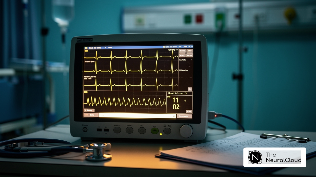
The peaked T waves meaning presents a diagnostic challenge on ECGs, often indicating serious underlying heart conditions. These waves are characterized by their tall, narrow, and symmetrical appearance, typically manifesting as sharp, pointed forms following the QRS complex. This shape indicates a notable change in how the ventricles repolarize, which is vital for diagnosing different heart conditions. Clinically, the peaked T waves meaning is often linked to hyperkalemia, characterized by a serum potassium level surpassing 5.5 mEq/L, and early myocardial ischemia, among other heart abnormalities. However, many healthcare professionals struggle to identify these critical features in ECG readings, which can signal serious underlying issues that need quick action.
Recent studies show that T shape morphology can be a key predictor of major adverse cardiovascular events (MACE) in patients with myocardial ischemia. For instance, T-wave characteristics, including onsets and offsets, have been shown to independently predict MACE, underscoring the need for meticulous ECG analysis. Furthermore, a systematic diagnostic approach that includes clinical assessment and laboratory tests is vital for identifying the causes of T-wave changes, ensuring timely and effective management.
Case studies demonstrate the clinical significance of elevated T shapes in heart diagnostics. For instance, the MaxYield platform enhances ECG analysis by concentrating on T characteristics, utilizing advanced noise filtering and signal recognition to improve diagnostic accuracy. MaxYield's beat-by-beat analysis capability allows for the processing of 200,000 heartbeats in under 5 minutes, empowering healthcare providers to make informed decisions. Identifying the peaked T waves meaning as vital signs of hyperkalemia enables swift action, possibly averting serious heart complications. Additionally, with FDA Class II SaMD clearance-pending status, MaxYield ensures compliance with regulatory standards, further enhancing its credibility. Overall, this delay in recognition can lead to severe consequences for patient health.

Explore Causes of Peaked T Waves: Physiological and Pathological Factors
The peaked T waves meaning in ECG readings can arise from various physiological and pathological factors, each carrying significant implications for heart health. Key causes include:
- Hyperkalemia: Elevated potassium levels in the blood are a primary contributor to peaked T waves. This condition often occurs in patients with renal failure or those on potassium-sparing diuretics, leading to notable alterations in cardiac repolarization.
- Myocardial Ischemia: In the initial phases of myocardial infarction, elevated T patterns can signify restricted blood flow to the heart muscle, acting as an essential warning signal for clinicians.
- Electrolyte Imbalances: Conditions such as hypomagnesemia and hypocalcemia can also result in T abnormalities, emphasizing the necessity for careful monitoring of electrolyte levels in patients.
- Cerebral Events: Although uncommon, elevated T forms may be linked to neurological disorders such as subarachnoid hemorrhage, highlighting the various elements that can affect ECG readings.
These conditions can complicate diagnosis and necessitate immediate clinical attention. By understanding these underlying causes, clinicians can enhance their diagnostic accuracy and improve patient care.

Analyze Diagnostic Techniques for Peaked T Waves: Testing and Interpretation
Diagnosing peaked T waves presents several challenges that require a systematic approach that incorporates several key diagnostic techniques:
- Electrocardiogram (ECG): The cornerstone for identifying peaked T waves, a 12-lead ECG offers a detailed view of the heart's electrical activity, enabling precise assessment of T wave morphology. Recent advancements in ECG technology, particularly with Neural Cloud Solutions' MaxYield™, have significantly enhanced diagnostic accuracy. MaxYield™ effectively analyzes ECG signals, filtering out interference to highlight key characteristics in each heartbeat. This allows for the identification of subtle variations in T patterns that could indicate heart issues. This progress is vital for health tech developers facing challenges in integrating advanced ECG solutions effectively.
- Clinical History: A comprehensive patient history is vital. Factors such as renal function, medication use, and symptoms of ischemia must be evaluated. It's important to consider medications like ACE inhibitors or potassium-sparing diuretics, as they can elevate T patterns during diagnosis. Symptoms related to elevated T patterns, including muscle weakness and heart issues, should also be observed as they can direct further assessment.
- Laboratory Tests: Blood tests that measure serum potassium levels and renal function are critical for confirming hyperkalemia or other electrolyte imbalances. Regular monitoring of these levels is crucial, especially in patients with risk factors for cardiac disturbances. Timely recognition of elevated T patterns can prevent severe cardiac complications, making it essential to understand the peaked T waves meaning as it relates to hyperkalemia being a primary factor for peaked T formations.
- Continuous Monitoring: In acute settings, continuous ECG monitoring is often necessary to track changes in T shape morphology over time, particularly in patients suspected of having myocardial ischemia. MaxYield™ supports this by delivering beat-by-beat analysis, outputting an analysis of 200,000 heartbeats in less than 5 minutes. This ongoing observation can provide insights into the progression of heart conditions and inform timely interventions.
Understanding these diagnostic tools is essential for effective patient management. Recognizing elevated T patterns in a timely manner can greatly impact treatment outcomes. As Dr. Alan Rabinowitz emphasizes, early and accurate detection of heart rhythm problems can mean the difference between life-saving intervention and a missed opportunity. With MaxYield™, healthcare professionals can enhance their diagnostic capabilities, ultimately improving patient outcomes.

Implement Treatment Strategies for Conditions Linked to Peaked T Waves
Effectively treating peaked T waves meaning involves navigating a complex landscape of underlying causes and treatment strategies. Key strategies include:
- Hyperkalemia Management: Immediate intervention often involves administering calcium gluconate to stabilize cardiac membranes, followed by insulin and glucose to facilitate the intracellular shift of potassium. In severe cases, dialysis may be necessary to effectively lower potassium levels.
- Ischemia Treatment: For patients undergoing myocardial ischemia, prompt revascularization via angioplasty or thrombolysis is essential, supplemented by antiplatelet therapy to improve blood flow and lessen heart strain.
- Electrolyte Correction: Adjusting any electrolyte imbalances through dietary changes or supplements is crucial for preventing the recurrence of elevated T forms, ensuring stable cardiac function.
- Monitoring and Follow-Up: Keeping a close eye on ECG readings and scheduling regular follow-ups is crucial for checking how well treatments are working and avoiding complications.
By employing these treatment strategies, healthcare professionals can effectively improve patient outcomes and decrease the risks related to the peaked T waves meaning. Furthermore, integrating Neural Cloud Solutions' MaxYield™ platform can enhance the efficiency of ECG analysis, allowing for automated labeling and data extraction that reduces operational costs. Expert endorsements from Dr. Alan Rabinowitz, Dr. Brett Heilbron, and Dr. Marc W. Deyell emphasize that MaxYield™ rivals human interpretation in precision and effectively reduces noise, thereby improving the overall quality of ECG assessments. This integration not only streamlines processes but also empowers healthcare providers to focus on critical decision-making, ultimately leading to better patient care. By leveraging advanced tools like MaxYield™, healthcare professionals can enhance their diagnostic capabilities and ultimately improve patient outcomes.

Conclusion
Healthcare professionals often struggle to accurately interpret ECG features, leading to potential misdiagnoses and delayed treatments. Peaked T waves indicate serious health issues like hyperkalemia and myocardial ischemia, which require immediate attention to prevent complications. By understanding T wave morphology, healthcare professionals can make timely decisions that enhance patient outcomes.
This article highlights the multifaceted nature of peaked T waves, examining their causes, diagnostic techniques, and treatment strategies. Key factors such as electrolyte imbalances, renal function, and the role of advanced diagnostic tools like MaxYield™ are emphasized as vital components in the assessment and management of these critical cardiac markers. Additionally, a systematic approach to diagnosing and treating conditions associated with peaked T waves is crucial for timely interventions that can save lives.
By prioritizing education and awareness around peaked T waves, the healthcare community can significantly reduce the risks associated with these ECG abnormalities.
Frequently Asked Questions
What are peaked T waves and why are they significant in ECG readings?
Peaked T waves are characterized by their tall, narrow, and symmetrical appearance, often indicating serious underlying heart conditions. They appear as sharp, pointed forms following the QRS complex and signify notable changes in ventricular repolarization, which is crucial for diagnosing various heart conditions.
What conditions are commonly associated with peaked T waves?
Peaked T waves are often linked to hyperkalemia, which is characterized by a serum potassium level exceeding 5.5 mEq/L, and early myocardial ischemia, among other heart abnormalities.
How can T wave morphology predict cardiovascular events?
Recent studies indicate that T wave morphology can be a key predictor of major adverse cardiovascular events (MACE) in patients with myocardial ischemia. Specific characteristics of T waves, including their onsets and offsets, have been shown to independently predict MACE.
What is the importance of a systematic diagnostic approach for T-wave changes?
A systematic diagnostic approach that includes clinical assessment and laboratory tests is essential for identifying the causes of T-wave changes, ensuring timely and effective management of potential heart conditions.
How does the MaxYield platform enhance ECG analysis?
The MaxYield platform improves ECG analysis by focusing on T wave characteristics, utilizing advanced noise filtering and signal recognition to enhance diagnostic accuracy. It offers beat-by-beat analysis, processing 200,000 heartbeats in under 5 minutes to support informed decision-making by healthcare providers.
How can identifying peaked T waves impact patient health?
Recognizing peaked T waves as critical indicators of hyperkalemia allows for swift intervention, potentially preventing serious heart complications and improving patient outcomes.
What is the regulatory status of the MaxYield platform?
The MaxYield platform is currently pending FDA Class II SaMD clearance, ensuring it meets regulatory standards and enhancing its credibility in the healthcare field.
List of Sources
- Define Peaked T Waves: Characteristics and Clinical Importance
- theneuralcloud.com (https://theneuralcloud.com/post/master-peaked-t-waves-essential-ecg-analysis-techniques)
- mdsearchlight.com (https://mdsearchlight.com/procedures/ecg-t-wave)
- Understanding Peaked T Waves (https://clinician.com/blogs/clinicians/understanding-peaked-t-waves)
- Explore Causes of Peaked T Waves: Physiological and Pathological Factors
- De Novo Hyperkalemia Increases Risks for Death, Hospitalizations (https://renalandurologynews.com/news/hyperkalemia-ups-risk-mortality-cardiovascular-events-hospitalizations-icu)
- healio.com (https://healio.com/cardiology/learn-the-heart/ecg-review/ecg-interpretation-tutorial/68-causes-of-t-wave-st-segment-abnormalities)
- Why T Waves Peak in Hyperkalemia (https://ecglectures.com/blog/peaked-t-waves-in-hyperkalemia)
- Unmasking Hyperkalemia: Highlighting Critical ECG Changes (https://powerfulmedical.com/blog/hyperkalemia-ecg-critical-changes)
- Analyze Diagnostic Techniques for Peaked T Waves: Testing and Interpretation
- Study Shows Promising Results for ECG Technology (https://dicardiology.com/content/study-shows-promising-results-ecg-technology)
- ECG T Wave - StatPearls - NCBI Bookshelf (https://ncbi.nlm.nih.gov/books/NBK538264)
- yourhealthmagazine.net (https://yourhealthmagazine.net/article/heart-disease-stroke-and-diabetes/peaked-t-waves-what-they-mean-for-your-heart-health)
- Implement Treatment Strategies for Conditions Linked to Peaked T Waves
- Hyperkalemia-Peaked T waves and ECG Recognition - Medcram Blog (https://blog.medcram.com/cme-courses/hyperkalemia-peaked-t-waves-and-ecg-recognition)
- Hyperkalemia Treatment & Management: Approach Considerations, Initial Emergency Management, Pharmacologic Therapy and Dialysis (https://emedicine.medscape.com/article/240903-treatment)
- Hyperkalemia – Diagnosis and Treatment : Emergency Care BC (https://emergencycarebc.ca/clinical_resource/clinical-summary/hyperkalemia-diagnosis-and-treatment)