Overview
Mastering EKG lead placement is essential for achieving accurate cardiac readings. Improper electrode positioning can result in misdiagnosis and delayed treatment. A comprehensive understanding of lead functionality, along with careful preparation of both the patient and the environment, greatly enhances signal fidelity. This improvement leads to better diagnostic outcomes, ultimately benefiting patient care.
Introduction
Mastering EKG lead placement is not merely a technical skill; it is an essential element in accurately assessing cardiac health. As healthcare professionals navigate the complexities of electrocardiograms, understanding the nuances of lead functionality and placement techniques becomes crucial. Even the most experienced practitioners may face challenges that can lead to misdiagnosis or delayed treatment. Therefore, identifying best practices for ensuring precise EKG readings is vital. Additionally, exploring how innovative technologies can enhance this critical process will further support healthcare professionals in their efforts.
Understand EKG Basics and Lead Functionality
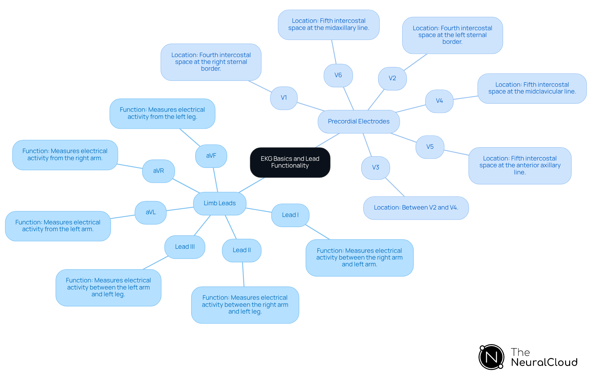
An electrocardiogram (EKG) records the heart's electrical signals via strategically positioned electrodes on the skin, with each connection providing a unique perspective of cardiac activity. The standard 12-lead EKG comprises:
- Limb Leads (I, II, III, aVR, aVL, aVF): Positioned on the arms and legs, these leads provide insights into the heart's electrical activity in the frontal plane, essential for diagnosing various cardiac conditions.
- Precordial Electrodes (V1 to V6): Positioned on the chest, these electrodes offer a perspective of the heart's activity in the horizontal plane. Each precordial electrode corresponds to specific anatomical locations, facilitating detailed analysis of cardiac function.
Comprehending these basics is essential for healthcare practitioners, as the quality of EKG signals is directly influenced by precise EKG lead placement. Variability in electrode positioning can significantly affect , such as those for left ventricular hypertrophy. Studies indicate that a substantial percentage of healthcare professionals utilize 12-lead EKG systems, underscoring the importance of mastering electrode functionality for effective interpretation.
Case studies have shown that correct electrode positioning enhances signal fidelity, minimizing noise and artifacts like muscle contractions and external electrical interference that can obscure true cardiac signals. The integration of Neural Cloud Solutions' MaxYield™ platform effectively addresses the challenges posed by these signal artifacts. MaxYield™ employs advanced noise filtering and distinct wave recognition, ensuring that even in recordings with high levels of noise, critical data is identified and labeled accurately. Furthermore, the algorithm evolves with each use, continuously improving its accuracy and efficiency in diagnostic yield.
Cardiologists emphasize that the operation of electrodes, specifically regarding EKG lead placement, is crucial in EKG interpretation, as misplacement can result in misdiagnosis and postponed treatment. Therefore, a thorough understanding of EKG basics is not just beneficial but essential for accurate cardiac assessments. Additionally, caution should be exercised regarding torso placement of limb leads, as this method should not be used interchangeably with standard ECGs for serial comparison, which can affect diagnostic accuracy.

Prepare the Patient and Environment for EKG Testing
Before conducting an EKG, it is essential to follow preparatory steps, including proper EKG lead placement, to ensure accurate results.
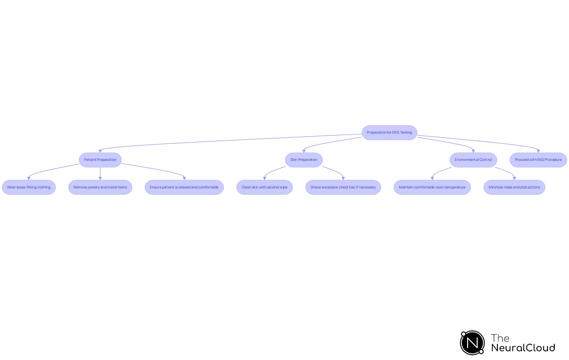
Patient Preparation: Instruct the patient to wear loose-fitting clothing that allows easy access to the chest, arms, and legs. Request the individual to remove any jewelry or metal items that might disrupt the sensors. Ensure the individual is relaxed and comfortable; anxiety can significantly impact heart rate and rhythm, potentially leading to inaccurate readings.
Skin Preparation: Clean the skin where electrodes will be placed using an alcohol wipe to remove oils and dirt, ensuring optimal electrode adhesion. If the individual has excessive chest hair, consider shaving the area to enhance electrode contact and signal clarity.
Environmental Control: Maintain a comfortable room temperature to prevent the patient from shivering, which can introduce artifacts in the EKG signal. Minimize noise and distractions to help the individual stay calm during the procedure. Research indicates that environmental factors, such as noise and temperature, can affect EKG results, making a controlled environment crucial for accurate readings. A serene testing environment is essential for obtaining reliable cardiac readings, as emphasized by healthcare professionals. Implementing best practices for environmental control not only enhances comfort for individuals but also contributes to the reliability of the EKG results.
Incorporating advanced technologies like Neural Cloud Solutions' MaxYield™ platform can further enhance EKG accuracy. This platform and data extraction, addressing challenges such as physiological variability and signal artifacts. By streamlining processes and reducing operational costs, the integration enables a more efficient analysis, ensuring improved outcomes for individuals. By following these preparatory steps and leveraging innovative solutions, healthcare professionals can significantly improve EKG lead placement results. After preparing the patient and environment, the next step is to proceed with the EKG procedure.

Execute Proper EKG Lead Placement Techniques
Follow these steps for accurate [EKG lead placement](https://theneuralcloud.com/post/master-ecg-artefacts-strategies-for-accurate-analysis-and-mitigation):
-
Limb Leads:
- Place the right arm (RA) electrode on the right wrist or forearm.
- Place the left arm (LA) sensor on the left wrist or forearm.
- Place the right leg (RL) sensor on the right ankle or lower leg.
- Place the left leg (LL) sensor on the left ankle or lower leg.
-
Precordial Leads:
- V1: Place in the 4th intercostal space at the right sternal border.
- V2: Place in the 4th intercostal space at the left sternal border.
- V3: Place midway between V2 and V4.
- V4: Place in the 5th intercostal space at the midclavicular line.
- V5: Place in the 5th intercostal space at the anterior axillary line.
- V6: Place in the 5th intercostal space at the midaxillary line.
Ensure that each conductor is securely attached and that the skin is properly prepared to minimize impedance during EKG lead placement.

Verify Lead Placement and Troubleshoot Common Issues
After placing the leads, follow these steps to verify and troubleshoot:
-
Verify Lead Placement:
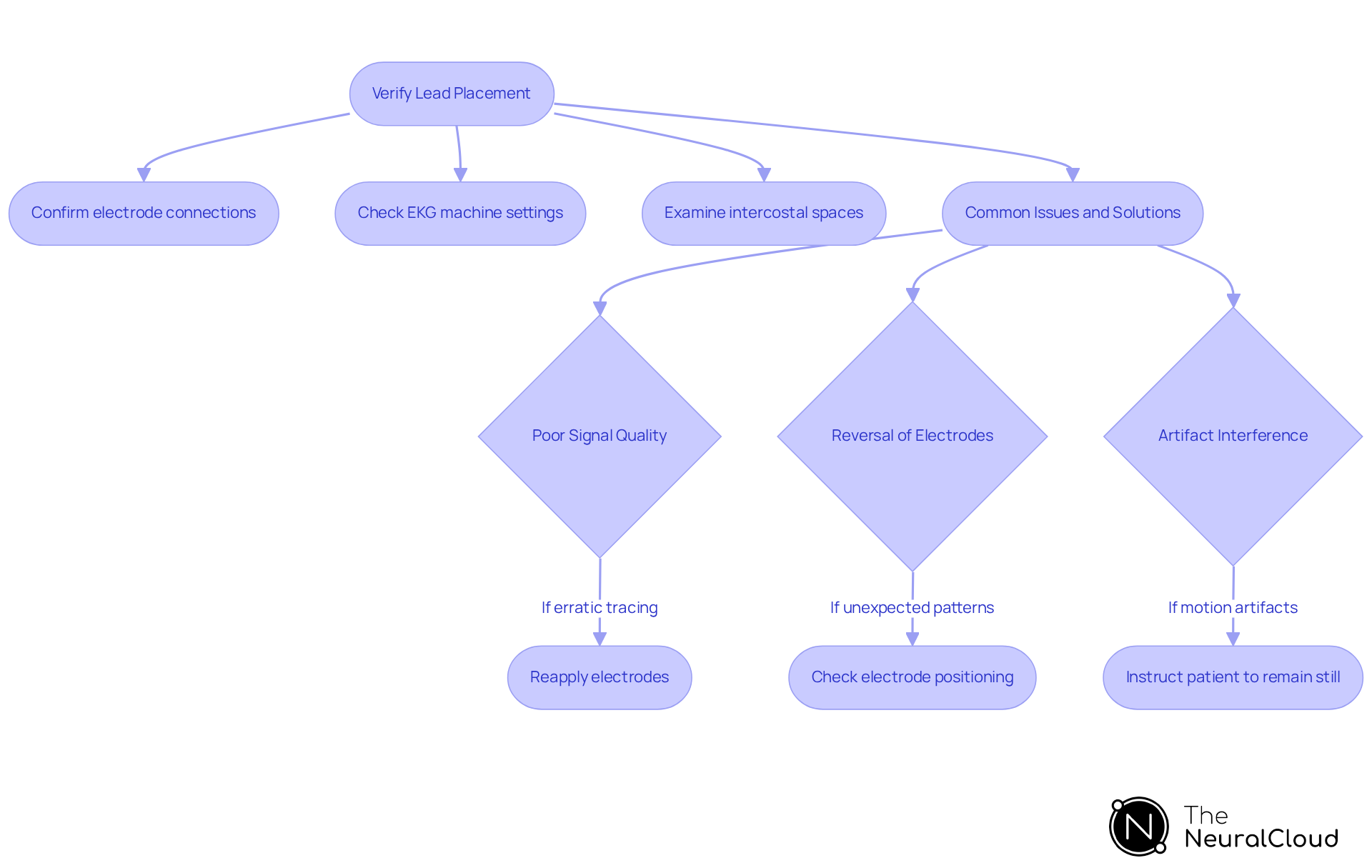
- Confirm that each lead is connected to the correct electrode and that the electrodes are securely attached to the skin. Proper connection is essential for precise measurements, as loose sensors can result in poor signal quality. Examining intercostal spaces is vital for accurate device placement, ensuring sensors are arranged to record genuine cardiac activity. The integration of wearable technology with Neural Cloud Solutions' MaxYield™ platform enhances this process by providing automated labeling and noise filtering, ensuring a clean signal for analysis while contributing to cost reduction by minimizing the need for manual adjustments.
- Ensure that the EKG machine is set to the correct lead configuration to avoid misinterpretation of the heart's electrical activity.
-
Common Issues and Solutions:
- Poor Signal Quality: If the EKG tracing appears erratic or unclear, check for loose electrodes or inadequate skin contact. Reapply electrodes as necessary, ensuring they are placed on fleshy areas while avoiding bones or scars to capture true cardiac activity. Utilizing MaxYield™ can automate the noise filtering process, significantly improving signal clarity and reliability, which reduces operational costs associated with retesting.
- Reversal of Electrodes: Unexpected patterns in the EKG may indicate that limb electrodes are swapped (e.g., right arm with left arm). Check electrode positioning promptly to rectify errors, as incorrect placement can greatly influence the precision of the EKG tracing. As noted by ljordan2101, 'Electrode placement directly affects the quality of the EKG tracing; improper placement can lead to inaccurate readings and artifacts,' reinforcing the importance of accurate EKG lead placement in relation to the capabilities of MaxYield™.
- Artifact Interference: To minimize artifacts, instruct the individual to remain still during the procedure and ensure no external electrical devices are nearby that could disrupt the readings. This is essential to prevent that can distort results. The advanced algorithms of MaxYield™ can assist in recognizing and filtering out these artifacts, enhancing the accuracy of the readings and contributing to overall efficiency.
By following these verification and troubleshooting steps, you can enhance the accuracy and reliability of EKG results, ultimately improving care for individuals. Documenting the EKG procedure is crucial as it provides a record of the findings and can impact ongoing patient care and treatment decisions.

Conclusion
Mastering EKG lead placement is a critical skill for healthcare professionals, as it directly impacts the accuracy of cardiac readings. Understanding the function of each lead, from limb to precordial electrodes, ensures that practitioners can effectively interpret the heart's electrical signals. Proper lead placement minimizes artifacts and enhances signal clarity, leading to more reliable diagnostic outcomes.
The article emphasizes the importance of thorough patient and environmental preparation, detailing steps such as:
- Ensuring skin cleanliness
- Maintaining a calm atmosphere
It outlines proper techniques for lead placement and verification, highlighting common pitfalls and their solutions. Utilizing advanced technology like Neural Cloud Solutions' MaxYield™ platform can further enhance the accuracy and efficiency of EKG readings, addressing challenges such as noise and electrode misplacement.
The MaxYield™ platform features advanced algorithms that optimize lead placement and signal processing. This technology improves ECG analysis by reducing noise interference and ensuring accurate electrode positioning, which is crucial for reliable cardiac assessments. By implementing these features, healthcare professionals benefit from enhanced diagnostic accuracy and improved patient care.
Ultimately, the significance of accurate EKG readings cannot be overstated. By implementing best practices and leveraging innovative solutions like the MaxYield™ platform, healthcare professionals can improve patient care and outcomes. A commitment to mastering EKG lead placement techniques is essential for delivering precise cardiac assessments and ensuring timely, effective treatment for patients.
Frequently Asked Questions
What is an electrocardiogram (EKG)?
An electrocardiogram (EKG) is a test that records the heart's electrical signals through electrodes placed on the skin, providing insights into cardiac activity.
What are the main components of a standard 12-lead EKG?
The standard 12-lead EKG consists of limb leads (I, II, III, aVR, aVL, aVF) positioned on the arms and legs, and precordial electrodes (V1 to V6) placed on the chest.
What do the limb leads measure?
Limb leads measure the heart's electrical activity in the frontal plane, which is essential for diagnosing various cardiac conditions.
What is the role of precordial electrodes in an EKG?
Precordial electrodes provide a perspective of the heart's activity in the horizontal plane, corresponding to specific anatomical locations for detailed cardiac function analysis.
Why is proper EKG lead placement important?
Proper EKG lead placement is crucial because it directly influences the quality of EKG signals, affecting diagnostic criteria and potentially leading to misdiagnosis if not done correctly.
How can incorrect electrode positioning affect EKG interpretation?
Incorrect electrode positioning can result in misdiagnosis and delayed treatment, making a thorough understanding of EKG basics essential for accurate cardiac assessments.
What challenges do signal artifacts pose in EKG recordings?
Signal artifacts such as muscle contractions and external electrical interference can obscure true cardiac signals, complicating interpretation.
How does Neural Cloud Solutions' MaxYield™ platform assist with EKG signal quality?
MaxYield™ employs advanced noise filtering and distinct wave recognition to enhance signal fidelity, ensuring critical data is accurately identified even in noisy recordings.
What caution should be taken regarding limb lead placement on the torso?
Limb leads should not be placed on the torso interchangeably with standard ECGs for serial comparison, as this can affect diagnostic accuracy.


