Introduction
Hyperacute T-waves are critical indicators of myocardial infarction, yet recognizing them poses significant challenges for healthcare professionals. The integration of advanced AI technologies empowers clinicians to enhance their diagnostic capabilities, facilitating more accurate and timely identification of these essential ECG changes.
Nevertheless, practitioners face the ongoing challenge of effectively navigating the complexities of hyperacute T-wave analysis while leveraging AI tools to optimize patient outcomes. This article explores the intricacies of hyperacute T-wave identification, the role of AI in ECG interpretation, and the essential criteria necessary to improve diagnostic accuracy in acute cardiac care.
Define Hyperacute T-Waves and Their Clinical Importance
are characterized by their morphology on an ECG, typically observed during the early stages of myocardial infarction. Their clinical significance is paramount, as these waves serve as indicators of acute coronary syndrome, necessitating immediate recognition and intervention. Research indicates that prompt identification of hyperacute T-waves can significantly influence treatment decisions, potentially reducing morbidity and mortality associated with acute coronary syndromes.
Recent studies emphasize that hyperacute T-wave patterns localize within the affected coronary distribution, often indicating ischemic injury in the area supplied by an occluded artery. For example, a multicenter study revealed that myocardial infarction was diagnosed in 445 out of 2457 patients, highlighting the importance of early detection. Additionally, the diagnostic efficacy of hyperacute T-wave amplitudes has shown that leads III, aVR, and V1 exhibit positive likelihood ratios, suggesting their potential utility in detecting acute coronary syndromes.
Cardiologists stress the importance of accurate diagnosis in emergency settings. As noted by specialists, understanding the nuances of these waveform changes is essential for healthcare providers, as it directly impacts management strategies in acute cardiac treatment. The ability to differentiate hyperacute T-wave from other T deflection irregularities, such as those associated with benign conditions, is crucial for effective treatment. This comparative approach enables clinicians to gain valuable insights into the underlying pathology, guiding interventions and optimizing patient outcomes.
Establish Criteria for Identifying Hyperacute T-Waves
To accurately identify hyperacute T-waves, clinicians should consider several key criteria:
- Morphology: Hyperacute T-waves are characterized by their significant height relative to the preceding R-wave, often exhibiting a pointed peak. When associated with occlusion myocardial infarction, the hyperacute T-wave is described as having a wide base and round peak, which is crucial for differentiation from other T-wave abnormalities. The AI technology enhances this analysis by effectively filtering noise, allowing for clearer identification of these critical morphological features.
- Duration: These transients typically appear suddenly and are generally brief, often enduring less than 0.2 seconds. This transient nature is a key feature in their identification, and MaxYield™'s algorithms can assist in tracking these fleeting events more efficiently.
- Lead Variability: It is essential to evaluate multiple leads, as hyperacute T-wave changes may be more prominent in certain leads, especially V2 and V3. MaxYield™'s advanced wave recognition can help clinicians quickly identify abnormalities, providing timely insights.
- Clinical Context: Correlating ECG findings with patient symptoms and history is vital. Hyperacute T-waveforms are frequently associated with acute coronary syndrome, such as chest pain, which can guide clinical decision-making. The adaptability of MaxYield™ allows for continuous improvement in diagnostic yield, ensuring that clinicians have the most accurate data at their disposal.
- Variants of Hyperacute T-Wave: Clinicians should also be aware of variations like the deWinter T wave and anterior QS waves with sharp T waves, which can assist in distinguishing these from other conditions. The integration of advanced analytics with MaxYield™ can further enhance the efficiency of identifying these variants, ultimately leading to cost reductions in management.
By following these guidelines and utilizing the features of MaxYield™, healthcare professionals can greatly improve diagnostic accuracy, resulting in prompt and efficient care management.

Explore Diagnostic Challenges and Considerations
Diagnosing abnormalities presents several challenges, primarily due to the influence of noise and artifacts.
Artifacts can significantly obscure T-wave morphology, leading to potential misinterpretation of patterns. Healthcare providers must remain vigilant in differentiating authentic wave alterations from artifacts caused by factors such as patient movement or equipment failure. Research indicates that baseline drift and muscle noise can hinder the recognition of wave morphology, possibly resulting in incorrect conclusions regarding a patient's cardiac condition.
Patient variability also plays a crucial role. Individual differences, including age, sex, and underlying health conditions, can markedly influence the morphology of the T-wave. Studies have shown that T-wave characteristics can vary across demographics, necessitating a nuanced understanding of these variations for accurate diagnosis. This variability emphasizes the importance of context within the broader clinical picture.
Furthermore, T-wave patterns may be misidentified as signs of other conditions, such as early repolarization or pericarditis. A thorough evaluation, incorporating correlation with medical history and further diagnostic tests, is essential to prevent misdiagnosis. Clinicians should be aware of common pitfalls, such as confusing T-wave changes with artifacts, which can occur in up to 4% of ECGs performed.
By recognizing these challenges, healthcare professionals can adopt a more careful and informed approach to ECG interpretation. This ultimately leads to improved accuracy and enhanced patient care.

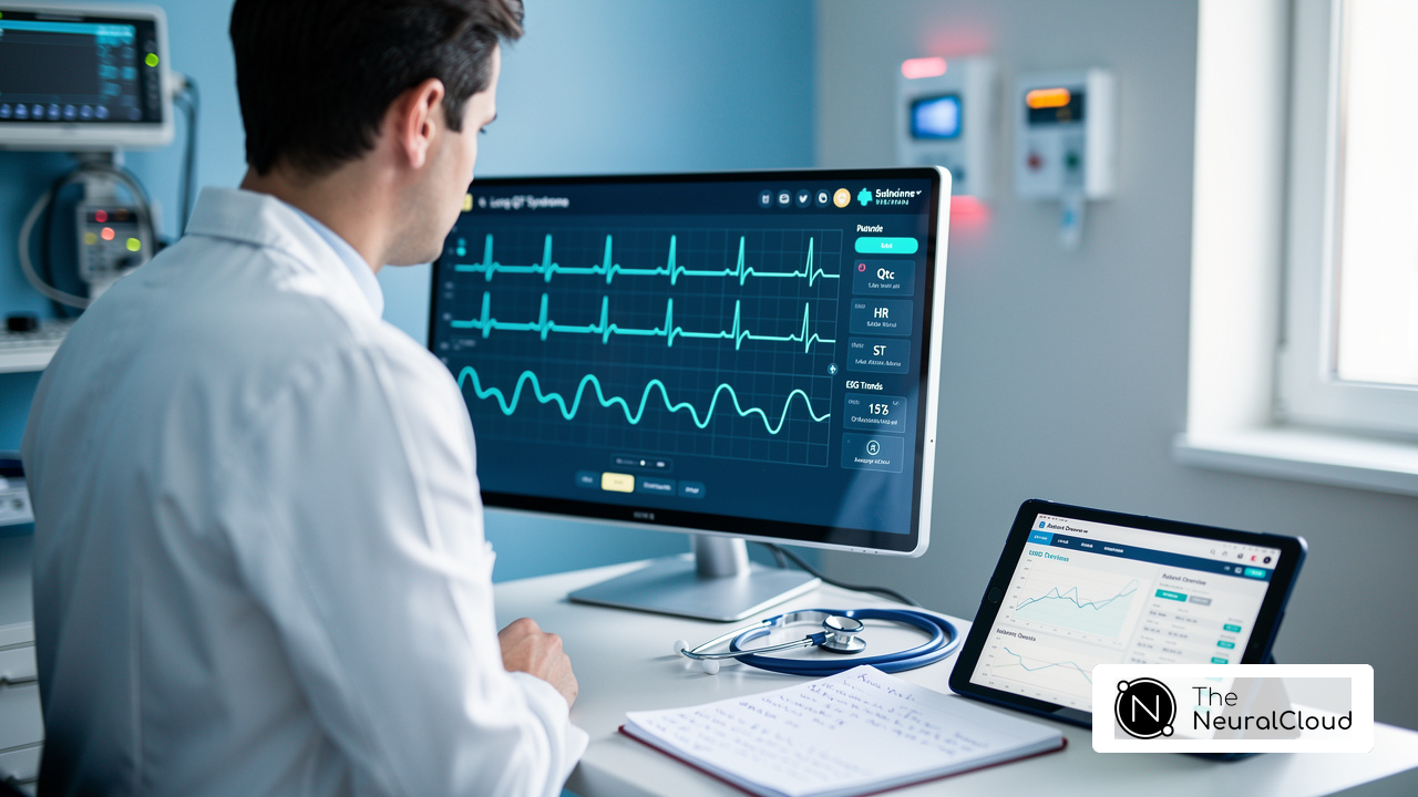
Integrate Advanced AI Technologies for Enhanced ECG Interpretation
Integrating advanced AI technologies into ECG interpretation significantly enhances the identification of hyperacute T-wave abnormalities through several key mechanisms.
- Automated Detection: Neural Cloud Solutions' system is designed to recognize patterns with remarkable accuracy. This automation alleviates the workload on clinicians, effectively minimizing human error and allowing for more reliable diagnostics.
- MaxYield™ can process ECG data in real-time, delivering immediate insights that empower healthcare professionals to make timely clinical decisions. This is crucial in acute care settings where every second counts, transforming noisy recordings into detailed insights and enabling beat-by-beat analysis of up to 200,000 heartbeats in less than 5 minutes.
- Continuous Learning: The AI model is built to evolve by learning from new data inputs. This aligns with the latest advancements in cardiology, enhancing the overall effectiveness of ECG interpretation.
Furthermore, statistics show that 79% of cardiac care providers have lost valuable clinical time due to incomplete or hard-to-access data, highlighting the necessity of AI in optimizing workflows. MaxYield™'s advanced analysis and wave recognition capabilities further illustrate the effectiveness of AI technologies, allowing for the identification of P-wave, QRS complex, and T-wave features that support confident clinical decisions.
As noted by experts in the field, AI technologies are transforming cardiac diagnostics. By embracing these AI technologies, healthcare professionals can streamline their workflows, improve diagnostic accuracy, and ultimately enhance patient outcomes, making a significant impact in the field of cardiac care.

Conclusion
Hyperacute T-waves serve as critical indicators of myocardial infarction, and their timely recognition can significantly influence patient outcomes. Understanding their morphology, duration, and clinical context is essential for healthcare professionals, as these factors directly impact diagnosis and management strategies in acute cardiac care. The integration of advanced AI technologies, such as MaxYield™, offers a transformative approach to enhance the accuracy and efficiency of ECG interpretation. This allows for real-time analysis and reduces the potential for human error.
The importance of hyperacute T-wave analysis cannot be overstated, as it highlights the need for clinicians to remain vigilant in identifying these patterns amidst potential diagnostic challenges. Factors such as signal artifacts and physiological variability can complicate recognition, making it imperative for healthcare providers to adopt a thorough and informed approach. Furthermore, the continuous learning capabilities of AI systems can assist clinicians in staying abreast of evolving diagnostic standards, ensuring they provide the best possible care.
In summary, the integration of AI in ECG analysis represents not just a technological advancement but a paradigm shift in diagnosing and managing cardiac conditions. By leveraging innovative tools like MaxYield™, healthcare professionals can enhance their diagnostic capabilities, ultimately leading to improved patient outcomes in cardiac care. The call to action is clear: embracing AI-driven solutions is essential for modern healthcare practices to ensure timely and effective responses to hyperacute T-wave abnormalities.
Frequently Asked Questions
What are hyperacute T-waves?
Hyperacute T-waves are characterized by their tall, peaked morphology on an ECG, typically observed during the early stages of myocardial infarction.
Why are hyperacute T-waves clinically significant?
They serve as critical indicators of impending cardiac events, necessitating immediate recognition and intervention, which can significantly influence treatment decisions and potentially reduce morbidity and mortality associated with acute coronary syndromes.
How do hyperacute T-waves relate to myocardial infarction?
Hyperacute T-wave patterns often localize within the affected coronary distribution, indicating ischemic injury in the area supplied by an occluded artery.
What does research indicate about the recognition of hyperacute T-waves?
Research shows that prompt identification of early cardiac wave alterations, such as hyperacute T-waves, can significantly impact treatment decisions and patient outcomes.
Which ECG leads are noted for their diagnostic efficacy in detecting myocardial infarction?
Leads III, aVR, and V1 exhibit positive likelihood ratios, suggesting their potential utility in detecting myocardial infarction.
What do cardiologists emphasize regarding hyperacute T-waves?
Cardiologists stress the importance of rapid T deflections in clinical practice, as understanding these waveform changes is essential for effective management strategies in acute cardiac treatment.
How can healthcare providers differentiate hyperacute T-waves from other T deflection irregularities?
It is crucial for healthcare providers to differentiate hyperacute T-waves from other T deflection irregularities by understanding their nuances, which aids in accurate diagnosis and effective treatment planning.
List of Sources
- Define Hyperacute T-Waves and Their Clinical Importance
- pubmed.ncbi.nlm.nih.gov (https://pubmed.ncbi.nlm.nih.gov/40892623)
- Hyperacute T-Waves: A Comprehensive Pattern Exploration (https://powerfulmedical.com/blog/hyperacute-t-waves)
- annemergmed.com (https://annemergmed.com/article/S0196-0644(22)01327-0/fulltext)
- link.springer.com (https://link.springer.com/article/10.1007/s11886-025-02217-8)
- Establish Criteria for Identifying Hyperacute T-Waves
- ECG Cases 21: Hyperacute T-waves and Occlusion MI (https://emergencymedicinecases.com/hyperacute-t-waves-occlusion-mi)
- Explore Diagnostic Challenges and Considerations
- pubmed.ncbi.nlm.nih.gov (https://pubmed.ncbi.nlm.nih.gov/40695024)
- pmc.ncbi.nlm.nih.gov (https://pmc.ncbi.nlm.nih.gov/articles/PMC6931710)
- mdpi.com (https://mdpi.com/2078-2489/10/2/35)
- theneuralcloud.com (https://theneuralcloud.com/post/10-key-artefact-ecg-issues-and-their-solutions)
- pmc.ncbi.nlm.nih.gov (https://pmc.ncbi.nlm.nih.gov/articles/PMC6891233)
- Integrate Advanced AI Technologies for Enhanced ECG Interpretation
- tctmd.com (https://tctmd.com/news/ai-ecg-model-outperforms-stemi-criteria-identifying-acs-patients-occlusions)
- AI-powered ECG model outperforms doctors in detecting hidden heart disease (https://news-medical.net/news/20250721/AI-powered-ECG-model-outperforms-doctors-in-detecting-hidden-heart-disease.aspx)
- Philips Launches ECG AI Marketplace to Enhance Early Cardiac Diagnosis (https://usa.philips.com/a-w/about/news/archive/standard/news/press/2025/philips-launches-ecg-ai-marketplace-to-enhance-early-cardiac-diagnosis.html)
- acc.org (https://acc.org/About-ACC/Press-Releases/2025/03/29/17/21/AI-Model-Shows-High-Accuracy)
- philips.com (https://philips.com/a-w/about/news/archive/standard/news/press/2025/philips-redefines-cardiac-care-with-ai-driven-solutions-at-esc-congress-2025.html)