Overview
The article provides a comprehensive guide for mastering six lead ECG placement, highlighting its significance for accurate cardiac diagnostics. It outlines specific anatomical landmarks for electrode placement and details a step-by-step process to ensure proper technique. Furthermore, it introduces advanced technology, such as the MaxYield™ platform, which enhances signal quality and diagnostic reliability. By addressing the challenges in ECG analysis, the article sets the stage for understanding how the MaxYield™ platform can improve the process.
The MaxYield™ platform offers several key features that enhance ECG analysis:
- It utilizes advanced algorithms to filter out noise, ensuring clearer signal interpretation.
- Additionally, the platform integrates seamlessly with existing systems, making it user-friendly for healthcare professionals.
These features collectively lead to improved diagnostic accuracy, ultimately benefiting patient care.
In conclusion, the MaxYield™ platform not only streamlines the ECG analysis process but also provides significant advantages for healthcare professionals. By improving signal quality and reliability, it allows for more accurate diagnoses, which is crucial in clinical settings. This guide serves as an essential resource for those looking to master ECG placement and leverage advanced technology for better patient outcomes.
Introduction
Understanding the intricacies of six lead ECG placement is essential for healthcare professionals who aim to enhance cardiac diagnostics. This advanced approach not only provides a comprehensive view of the heart's electrical activity but also significantly improves the accuracy of detecting conditions such as atrial fibrillation. However, mastering the precise placement of electrodes presents a challenge; even minor errors can lead to misdiagnosis and compromised patient care.
How can professionals ensure they execute this critical procedure with the utmost accuracy and reliability?
Understand the Basics of Six Lead ECG Placement
The six lead ECG placement, or 6-lead electrocardiogram, captures the heart's electrical activity through six strategically placed electrodes on the body. This approach greatly enhances diagnostic capabilities by providing a more comprehensive view of the heart's electrical signals compared to conventional electrode placements. Understanding the fundamental principles of these components and their specific roles is crucial for effective cardiac diagnostics.
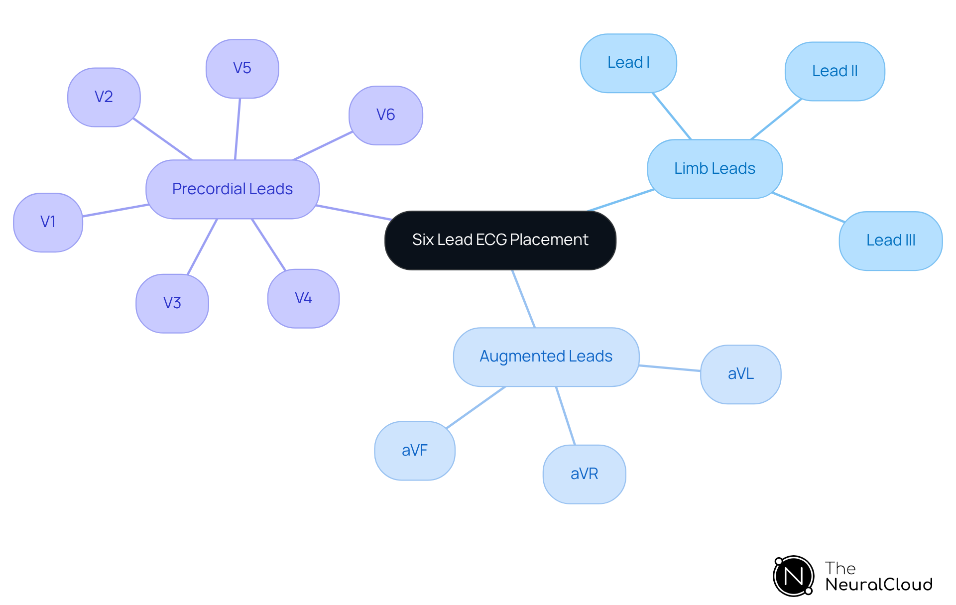
Each lead offers a unique perspective of the heart, enabling healthcare professionals to detect irregularities in rhythm and function with greater accuracy. The leads are categorized into:
- Limb leads (I, II, III)
- Augmented leads (aVR, aVL, aVF)
- Precordial leads (V1-V6)
This configuration provides detailed insights into the heart's activity, which is especially advantageous in identifying conditions such as atrial fibrillation (AF).
Recent studies reveal that the six lead ECG placement exhibits compared to single-lead ECGs, particularly in populations with frequent premature contractions. Notably, a study indicated that 75% of healthcare professionals favored the six-lead device over traditional 12-lead systems, citing ease of use and enhanced patient satisfaction. Furthermore, the handheld six-lead device has demonstrated a high negative predictive value of 99.8% at a QTc interval threshold of 500 milliseconds, reinforcing its effectiveness in clinical practice.
Integrating Neural Cloud Solutions' MaxYield™ platform with six lead ECG placement can further enhance analysis efficiency. MaxYield™ utilizes advanced noise filtering and distinct wave recognition to isolate and label critical data, even in recordings with high noise levels and artifacts. It provides beat-by-beat analysis, processing up to 200,000 heartbeats in less than 5 minutes, enabling healthcare professionals to convert noisy recordings into detailed insights. Familiarizing oneself with the fundamentals of six lead ECG placement, along with the application of MaxYield™, is essential for healthcare professionals aiming to enhance their diagnostic precision and improve patient outcomes.

Identify Key Anatomical Landmarks for Lead Placement
Precise sensor placement is essential for obtaining reliable ECG readings, and the MaxYield™ platform enhances this process through advanced noise filtering and wave recognition capabilities. To achieve optimal results, identifying key anatomical landmarks on the patient's body is crucial. The following guidelines outline the proper six-lead ECG placement:
- Right Arm (RA): Position the sensor on the right wrist or just below the right clavicle.
- Left Arm (LA): Position the sensor on the left wrist or just below the left clavicle.
- Left Leg (LL): Attach the sensor to the left ankle or just above the left hip.
- Right Leg (RL): Position the sensor on the right ankle or just above the right hip.
- V1: Place the device in the fourth intercostal space at the right sternal border.
- V2: Place the device in the fourth intercostal space at the left sternal border.
- V3: Position the sensor midway between V2 and V4.
- V4: Place the sensor in the fifth intercostal space at the midclavicular line.
- V5: Position the device in the fifth intercostal space at the anterior axillary line.
- V6: Position the lead in the fifth intercostal space at the midaxillary line.
Research indicates that even a 2-centimeter misplacement of electrodes can significantly distort ECG morphology, potentially leading to misdiagnosis. For instance, improper positioning of V1 and V2 can mimic anterior myocardial infarction (MI) and result in T-wave inversion. Thus, ensuring is vital for accurate diagnosis and effective patient care.
Healthcare professionals stress the significance of these anatomical points, emphasizing that proper six lead ECG placement can expedite the identification of critical conditions such as STEMI. By accurately recognizing these landmarks, you ensure that the sensors are correctly positioned, which is essential for obtaining reliable ECG readings. Furthermore, the MaxYield™ platform's ability to filter noise and recognize distinct waves enhances the clarity of the readings, facilitating improved diagnostic outcomes. It is also imperative to double-check that all electrodes are affixed in the correct locations to prevent discrepancies in the readings. Remember, it takes less than 30 seconds to determine the correct position for a 12-lead ECG, making efficiency a crucial factor in the process.

Follow Step-by-Step Instructions for Accurate Lead Placement
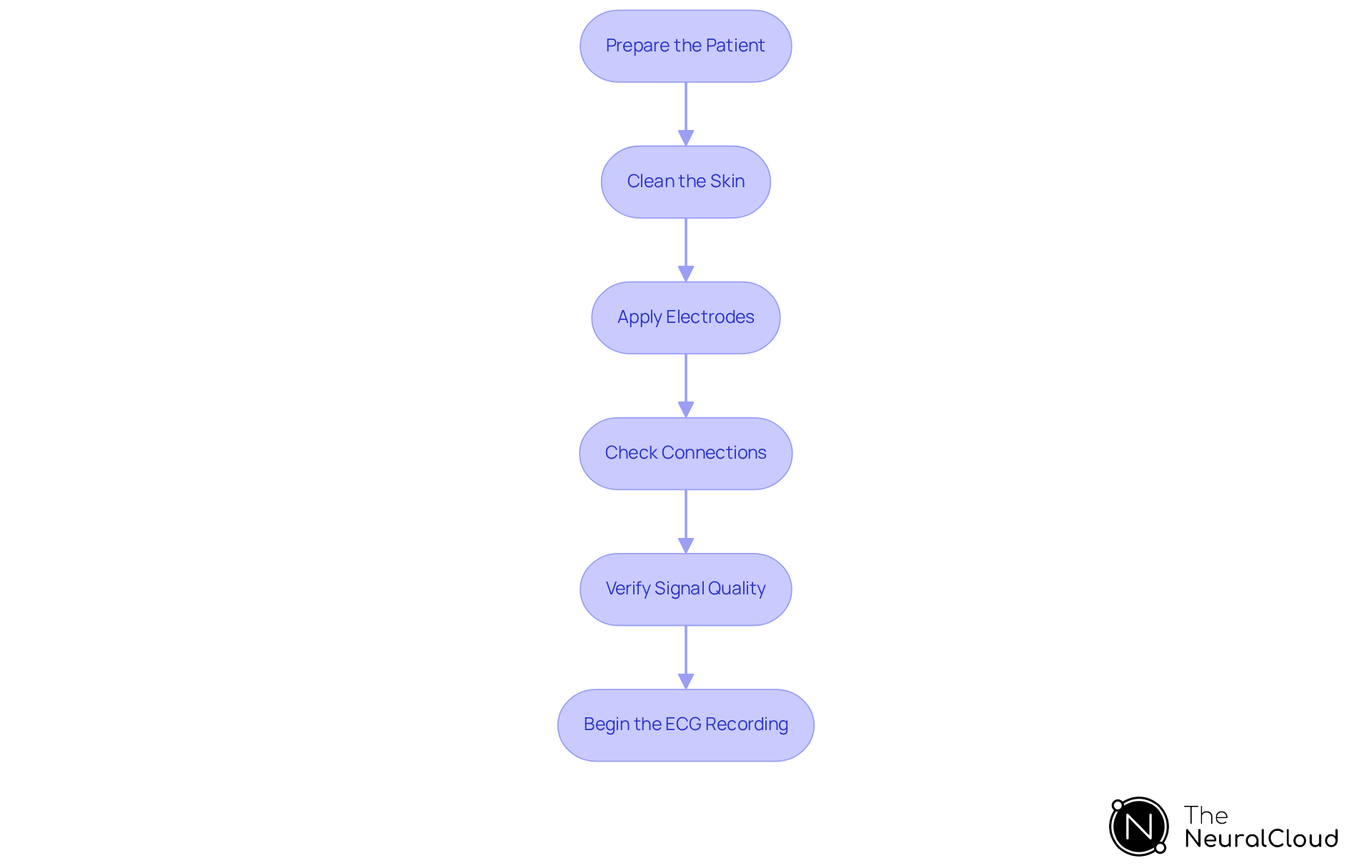
To ensure accurate placement of the six ECG leads, adhere to the following step-by-step instructions:
- Prepare the Patient: Position the patient comfortably, ideally lying down, and explain the procedure to alleviate any anxiety.
- Clean the Skin: Utilize alcohol wipes to cleanse the skin where sensors will be applied. This step is crucial as it reduces impedance and enhances signal quality, with studies indicating that significantly improves ECG data reliability.
- Apply Electrodes:
- Begin with the limb leads: Attach the Right Arm (RA), Left Arm (LA), Right Leg (RL), and Left Leg (LL) electrodes as previously identified.
- Proceed to the precordial electrodes: Position V1 and V2 first, followed by V3, V4, V5, and V6 in their assigned locations.
- Check Connections: Confirm that all sensors are securely attached and that the wires are properly connected to the ECG machine.
- Verify Signal Quality: Before initiating the ECG recording, assess the signal quality on the monitor. Look for clear waveforms devoid of excessive noise or artifacts. Research shows that maintaining low motion artifact levels is essential for accurate readings, particularly with Ag/AgCl electrodes, which perform well even without additional gel. The integration of Neural Cloud Solutions' MaxYield™ platform enhances this process by utilizing advanced noise filtering and distinct wave recognition, ensuring that critical data is accurately identified and labeled, even in challenging recording conditions.
- Begin the ECG Recording: Once all components are in place and verified, commence the ECG recording. Continuously monitor the process to ensure that the connections remain secure and that signal quality is maintained.
By following these steps, you can achieve accurate electrode positioning for six lead ECG placement, which is essential for obtaining trustworthy ECG results. Incorrect positioning of sensors can result in inaccurate readings, as highlighted by ECG specialists who stress the significance of proper wire application to avoid misdiagnosis. Case studies have demonstrated that adherence to these best practices not only enhances signal quality but also improves patient outcomes, particularly in settings where timely cardiac diagnostics are critical. The MaxYield™ platform's ability to salvage previously obscured sections of lengthy recordings further underscores its value in enhancing ECG analysis efficiency.

Troubleshoot Common Issues in Six Lead ECG Placement
Even with careful preparation, challenges can arise during the process of six lead ECG placement. Below are some common problems and their corresponding solutions:
- Poor Signal Quality: Noisy or unclear ECG signals often arise from insufficient connections. Ensure that the sensors are securely attached and re-clean the skin if necessary to enhance contact. Utilizing Neural Cloud Solutions' can significantly enhance signal clarity, as it employs advanced noise filtering to salvage previously obscured sections of recordings.
- Electrode Movement: If a connector becomes loose or dislodged, promptly reposition it and verify the signal quality again. Regular checks during the recording can prevent this issue. The MaxYield™ technology can help in quickly identifying and labeling critical data, even in recordings with movement artifacts.
- Patient Movement: Instruct the patient to remain still during the recording. If movement occurs, pause the recording and restart once the patient is settled. Research shows that patient movement can greatly affect signal quality, with a sensitivity of 57% and a specificity of almost 100% for detecting interchange between left arm and left leg sensors, resulting in misinterpretations. MaxYield™ can assist in isolating ECG waves from recordings affected by such artifacts, ensuring more reliable results.
- Skin Preparation Issues: Oily or dirty skin can hinder electrode adhesion. Ensure thorough cleaning with an alcohol pad or soap and water, and consider using conductive gel to enhance contact. Proper skin preparation is crucial for maximizing the effectiveness of the MaxYield™ platform, which thrives on clear signal input.
- Electrode Misplacement: Abnormal ECG results may suggest electrode misplacement. Verify the positioning of the markers against anatomical landmarks to ensure precision. Research indicates that nurses misplace ECG connections in up to 64% of all instances, and fewer than 20% of cardiologists accurately position V1 and V2 precordial electrodes, emphasizing the significance of correct technique. The MaxYield™ platform's AI-driven automation can help in quickly identifying misplacements and improving overall accuracy.
As Mahin Rehman, a specialist in Internal Medicine, states, 'Proper chest electrode positioning is crucial for an accurate ECG recording and its interpretation.' By being aware of these common issues and their solutions, and leveraging the capabilities of MaxYield™, you can enhance the accuracy and reliability of your recordings for six lead ECG placement. Furthermore, the economic burden of ECG mispositioning is significant, with an estimated cost of $3.2 billion annually due to unnecessary cardiovascular testing resulting from misinterpretations. MaxYield™ can help mitigate these costs by improving lead placement accuracy and reducing the need for repeat tests.

Conclusion
Mastering six lead ECG placement is essential for healthcare professionals seeking to enhance their diagnostic capabilities. This guide underscores the significance of precise electrode positioning, which markedly improves the accuracy of cardiac assessments. By grasping the fundamental principles and techniques of six lead ECG placement, practitioners can deliver more reliable diagnoses and enhance patient outcomes.
The article highlights key points, including:
- The unique perspectives provided by each lead
- The crucial role of anatomical landmarks in achieving accurate placements
- The step-by-step instructions necessary for effective execution
The integration of advanced technologies such as Neural Cloud Solutions' MaxYield™ platform significantly enhances the reliability of ECG readings by filtering noise and improving data clarity. Furthermore, common issues and troubleshooting strategies are discussed, ensuring that practitioners are well-equipped to address potential challenges during the ECG process.
Ultimately, the importance of accurate six lead ECG placement cannot be overstated. It transcends being merely a technical skill; it is a vital component of effective patient care that can facilitate timely interventions and better health outcomes. Healthcare professionals are encouraged to adopt these best practices and leverage available technologies to refine their techniques, thereby ensuring they deliver the highest standard of cardiac diagnostics.
Frequently Asked Questions
What is a six lead ECG placement?
A six lead ECG placement, or 6-lead electrocardiogram, captures the heart's electrical activity through six strategically placed electrodes on the body, providing a more comprehensive view of the heart's electrical signals compared to conventional electrode placements.
What are the different types of leads in a six lead ECG?
The leads in a six lead ECG are categorized into three types: Limb leads (I, II, III), Augmented leads (aVR, aVL, aVF), and Precordial leads (V1-V6).
How does the six lead ECG improve diagnostic capabilities?
The six lead ECG enhances diagnostic capabilities by offering unique perspectives of the heart, allowing healthcare professionals to detect irregularities in rhythm and function with greater accuracy.
What advantage does the six lead ECG have in detecting atrial fibrillation (AF)?
The six lead ECG placement provides detailed insights into heart activity, which is particularly advantageous for identifying conditions such as atrial fibrillation (AF). It has been shown to have superior diagnostic value for AF detection compared to single-lead ECGs.
What do recent studies indicate about healthcare professionals' preferences for ECG devices?
Recent studies indicate that 75% of healthcare professionals favored the six-lead device over traditional 12-lead systems, citing ease of use and enhanced patient satisfaction.
What is the negative predictive value of the handheld six-lead device?
The handheld six-lead device has demonstrated a high negative predictive value of 99.8% at a QTc interval threshold of 500 milliseconds, reinforcing its effectiveness in clinical practice.
How can the MaxYield™ platform enhance the analysis of six lead ECGs?
The MaxYield™ platform integrates with six lead ECG placement to enhance analysis efficiency by utilizing advanced noise filtering and distinct wave recognition, enabling beat-by-beat analysis and processing up to 200,000 heartbeats in less than 5 minutes.
Why is it important for healthcare professionals to understand six lead ECG placement and MaxYield™?
Familiarizing oneself with the fundamentals of six lead ECG placement and the application of MaxYield™ is essential for healthcare professionals aiming to enhance their diagnostic precision and improve patient outcomes.


