Overview
This article presents a step-by-step tutorial for analyzing ventricular tachycardia (VT) rhythm strips, highlighting the systematic approach necessary for accurate interpretation. It details specific analysis techniques, such as:
- Assessing heart rate
- Evaluating rhythm regularity
- Examining QRS complexes
Furthermore, it introduces advanced AI tools like MaxYield™, which enhance diagnostic accuracy and efficiency in clinical settings. By utilizing MaxYield™, healthcare professionals can significantly improve their ECG analysis capabilities, addressing common challenges in the interpretation of VT rhythms.
Introduction
Ventricular tachycardia (VT) poses a significant challenge in cardiac health, marked by a dangerously rapid heart rhythm that can result in severe complications if not addressed promptly. This tutorial explores the complexities of analyzing VT rhythm strips, providing healthcare professionals with step-by-step techniques to improve their diagnostic skills.
The integration of advanced tools and AI technology has made it more accessible than ever to enhance accuracy and efficiency in identifying VT. Mastering these techniques can not only transform individual patient outcomes but also redefine the standards of cardiac care.
Define Ventricular Tachycardia: Key Characteristics and Mechanisms
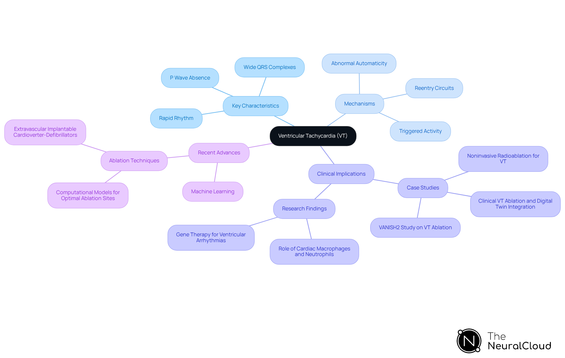
Ventricular tachycardia (VT) is characterized by a rapid heart rhythm originating from the ventricles, which can be identified on a vtach rhythm strip as three or more consecutive beats at a rate exceeding 100 beats per minute. The key characteristics of the vtach rhythm strip include wide QRS complexes (≥120 ms) that exhibit bizarre morphology, indicating abnormal ventricular depolarization. In most instances, P waves are not discernible, suggesting that atrial contractions are not synchronized with ventricular activity. The rhythm is often regular, although certain types of VT may present with irregular patterns.
The mechanisms underlying VT can be illustrated through a vtach rhythm strip, which may involve reentry circuits, triggered activity, and abnormal automaticity, often stemming from various cardiac conditions such as ischemic heart disease or cardiomyopathy. Recent studies have highlighted the role of machine learning algorithms in differentiating wide QRS complex tachycardias by analyzing QRS polarity direction and shifts. These algorithms demonstrate strong diagnostic accuracy when integrating features from both wide QRS tachycardia and baseline electrocardiograms.
Neural Cloud Solutions' MaxYield™ platform enhances this analysis by employing advanced noise filtering and wave recognition techniques. This allows for the , even in recordings with significant noise and artifacts. The efficiency of ECG analysis is improved, aiding in the identification of cardiac events and supporting confident clinical decision-making.
Furthermore, the VANISH2 study has strengthened the effectiveness of VT ablation in individuals with ischemic cardiomyopathy, providing strong evidence for enhanced clinical outcomes as indicated by the vtach rhythm strip. As noted by Gregory B. Lim, "Extravascular implantable cardioverter-defibrillators are safe and effective at detecting and terminating ventricular arrhythmias induced at the time of implantation," underscoring the importance of effective treatment options.
Case studies have shown that the integration of computational models can identify optimal ablation sites for treating infarct-related VT, which can be validated by the vtach rhythm strip, thereby eliminating the need for invasive electrical mapping. This advancement streamlines the treatment process and improves safety and outcomes for individuals. Additionally, research indicates that cardiac macrophages can protect against arrhythmias, while cardiac neutrophils may increase the likelihood of arrhythmia following myocardial infarction, underscoring the complex interplay of cellular mechanisms observed in a vtach rhythm strip.

Explore Clinical Implications of VT: Diagnosis and Patient Management
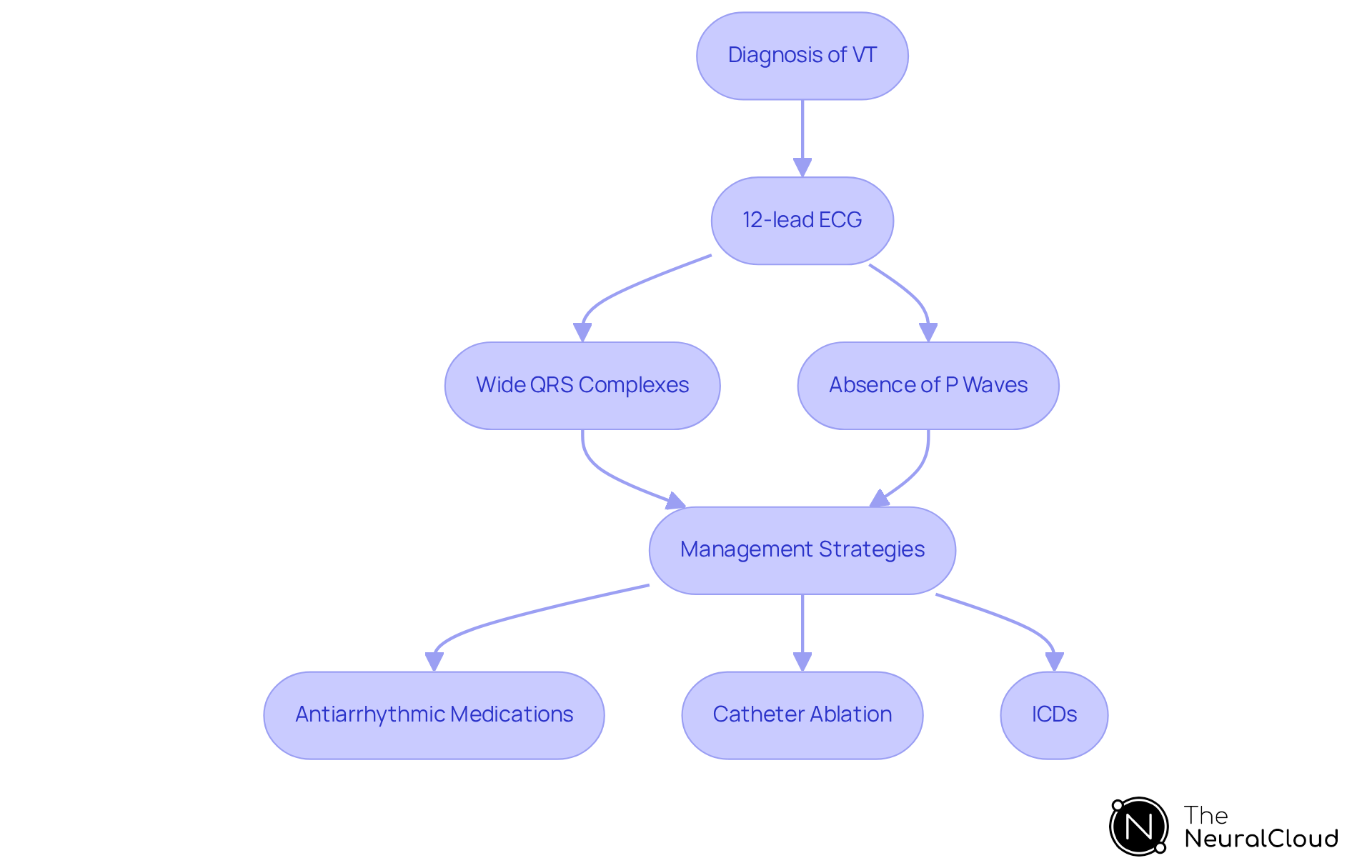
Ventricular tachycardia (VT) presents significant clinical risks, including syncope, heart failure, and sudden cardiac death. Accurate diagnosis is crucial and typically relies on a 12-lead ECG, where wide QRS complexes and the absence of P waves serve as key indicators of VT. The diagnostic accuracy for VT is notably high, with approximately 80% of wide-complex tachycardias ultimately diagnosed as VT, particularly in patients over 35 years old.
The 'Neural Cloud Solutions' platform enhances this diagnostic process by leveraging advanced noise filtering and distinct wave recognition capabilities. It effectively isolates ECG waves from recordings affected by baseline wander, movement, and muscle artifacts. MaxYield™ ensures that critical data is identified and labeled, even in challenging conditions. This continuous learning model evolves with each use, improving accuracy and efficiency over time, which is essential for managing VT effectively.
Management strategies for VT are multifaceted, potentially including:
- Antiarrhythmic medications
- Catheter ablation
- The use of implantable cardioverter-defibrillators (ICDs) for individuals at elevated risk of life-threatening arrhythmias
Notably, the combination of beta-blockers and amiodarone has been shown to be superior to beta-blockers alone in managing VT. The latest guidelines suggest a personalized approach, emphasizing the significance of risk stratification for sudden cardiac death and collaborative decision-making with individuals regarding ICD options, considering both quality of life and longevity.
Ongoing observation is essential for individuals with a history of VT, especially in acute care environments, to guarantee prompt identification and action during episodes. By integrating wearable technology with their system, healthcare providers can automate labeling and reduce expenses, thereby enhancing patient safety and improving overall management outcomes. This proactive approach allows healthcare providers to .

Analyze VT Rhythm Strips: Step-by-Step ECG Interpretation Techniques
Analyzing vtach rhythm strip necessitates a systematic approach, and the integration of MaxYield™ significantly enhances this process. To effectively assess the situation, follow these steps:
- Assess the Rate: Count the number of QRS complexes in a 6-second strip and multiply by 10 to determine the heart rate. A vtach rhythm strip typically indicates a ventricular tachycardia (V-Tach) rate that exceeds 100 beats per minute, which can considerably reduce cardiac output and elevate the risk of syncope or cardiac arrest. This system automates the counting process, delivering rapid and precise heart rate assessments.
- Evaluate the Rhythm: Determine if the rhythm is regular or irregular by measuring the distance between R waves. V-Tach often presents as a regular vtach rhythm strip, which is crucial for timely intervention. The system enhances rhythm evaluation through beat-by-beat analysis, facilitating quick identification of rhythm patterns.
- Examine the QRS Complex: Look for wide QRS complexes (≥120 ms) and assess their morphology for uniformity. A wide QRS indicates that the impulse originates from the ventricles, a key characteristic of V-Tach. This system provides detailed insights into QRS morphology, aiding in precise diagnosis.
- Identify P Waves: Determine if P waves are present; in V-Tach, they are typically absent, a key distinguishing feature. The platform's advanced algorithms assist in isolating and labeling these features effectively.
- Measure the PR Interval: In the vtach rhythm strip, the PR interval is usually not applicable due to the absence of P waves, further confirming the diagnosis. The system streamlines this measurement process, ensuring clarity in interpretation.
- Document Findings: Record your observations and compare them with established criteria for V-Tach diagnosis. This documentation is essential for effective management and treatment decisions, as rapid recognition of arrhythmias can significantly impact patient outcomes. The system facilitates seamless documentation and data management, enhancing clinical decision-making.
Understanding these steps is vital for accurate rhythm recognition, particularly in emergency situations where continuous practice and simulation training can refine skills and improve response effectiveness. With this solution, healthcare professionals can , leveraging automated, scalable, and AI-driven insights for enhanced clarity and efficiency.

Utilize Advanced ECG Analysis Tools: Enhancing Accuracy with AI Technology
Advanced ECG analysis tools, particularly those powered by AI, present significant improvements in the accuracy and efficiency of diagnosing a vtach rhythm strip. The challenges in ECG analysis often stem from noise interference, manual interpretation, and the need for continuous adaptation to diverse patient populations. The MaxYield™ platform addresses these challenges with a range of innovative features.
- Noise Reduction: MaxYield™ utilizes advanced noise filtering techniques, part of our Gold Standard Methodologies, to effectively eliminate noise and artifacts from ECG signals. This results in , enhancing the reliability of diagnostic outcomes.
- Automated Analysis: The platform automates the identification and labeling of key features in vtach rhythm strips. This automation allows clinicians to focus on interpretation rather than manual analysis, thus streamlining workflow and improving efficiency in clinical settings.
- Continuous Learning: By employing a continuous learning model, AI systems like MaxYield™ can learn from new data. This capability enhances their diagnostic abilities over time and allows them to adapt to various patient populations, which is crucial for improving accuracy across diverse clinical environments.
- Integration with Clinical Workflows: These advanced tools can seamlessly integrate into existing clinical systems. This integration enhances workflow efficiency and reduces operational costs, ultimately revolutionizing the ECG analysis process. Testimonials from healthcare professionals underscore the transformative impact of MaxYield™ on their diagnostic processes, highlighting its effectiveness in real-world applications.

Conclusion
Ventricular tachycardia (VT) presents a critical challenge in cardiac care, characterized by a rapid heart rhythm that can lead to severe complications if not properly diagnosed and managed. This article has provided a comprehensive overview of VT, detailing its key characteristics, underlying mechanisms, and the importance of accurate rhythm strip analysis. By leveraging advanced tools and techniques, healthcare professionals can enhance their diagnostic capabilities and improve patient outcomes.
The discussion highlighted the significance of recognizing wide QRS complexes and the absence of P waves on a VT rhythm strip as fundamental indicators of VT. It also emphasized the role of innovative platforms like MaxYield™ in facilitating accurate diagnosis through noise reduction and automated analysis. The MaxYield™ platform improves ECG analysis by offering features such as enhanced signal clarity, real-time interpretation, and user-friendly interfaces. These advantages lead to more accurate diagnoses, ultimately benefiting patient care.
Furthermore, management strategies for VT, including the use of antiarrhythmic medications and catheter ablation, were explored, underscoring the necessity of a personalized approach to treatment. As technology continues to advance, integrating AI-driven tools into clinical practice will not only streamline workflows but also enhance the accuracy of diagnoses. Embracing these innovations can empower healthcare providers to respond adeptly to the complexities of VT, ultimately safeguarding patient health and advancing cardiac care.
In conclusion, mastering VT rhythm strip analysis is essential for effective patient management and improved clinical outcomes. By utilizing advanced tools like MaxYield™, healthcare professionals can navigate the challenges of ECG analysis more effectively, leading to better patient outcomes and a deeper understanding of ventricular tachycardia.
Frequently Asked Questions
What is ventricular tachycardia (VT)?
Ventricular tachycardia (VT) is a rapid heart rhythm originating from the ventricles, characterized by three or more consecutive beats at a rate exceeding 100 beats per minute.
What are the key characteristics of a VT rhythm strip?
Key characteristics of a VT rhythm strip include wide QRS complexes (≥120 ms) with bizarre morphology, indicating abnormal ventricular depolarization, and the absence of discernible P waves, suggesting that atrial contractions are not synchronized with ventricular activity.
How does the rhythm of VT typically present?
The rhythm of VT is often regular, although certain types may present with irregular patterns.
What mechanisms underlie ventricular tachycardia?
The mechanisms underlying VT can include reentry circuits, triggered activity, and abnormal automaticity, often associated with cardiac conditions such as ischemic heart disease or cardiomyopathy.
How do machine learning algorithms assist in diagnosing VT?
Machine learning algorithms assist in diagnosing VT by analyzing QRS polarity direction and shifts, demonstrating strong diagnostic accuracy when integrating features from wide QRS tachycardia and baseline electrocardiograms.
What role does the MaxYield™ platform play in ECG analysis?
The MaxYield™ platform enhances ECG analysis by employing advanced noise filtering and wave recognition techniques, allowing for rapid isolation and labeling of critical ECG data, even in noisy recordings.
What findings did the VANISH2 study reveal about VT ablation?
The VANISH2 study provided strong evidence that VT ablation is effective in individuals with ischemic cardiomyopathy, leading to enhanced clinical outcomes.
What is the significance of extravascular implantable cardioverter-defibrillators in treating VT?
Extravascular implantable cardioverter-defibrillators are safe and effective at detecting and terminating ventricular arrhythmias, particularly at the time of implantation.
How can computational models aid in the treatment of infarct-related VT?
Computational models can identify optimal ablation sites for treating infarct-related VT, which can be validated by the VT rhythm strip, thus eliminating the need for invasive electrical mapping.
What is the role of cardiac macrophages and neutrophils in arrhythmias?
Cardiac macrophages can protect against arrhythmias, while cardiac neutrophils may increase the likelihood of arrhythmia following myocardial infarction, highlighting the complex interplay of cellular mechanisms in VT.


