Introduction
Recognizing hyperacute T waves on an ECG can significantly impact the early detection of myocardial infarction. However, many healthcare professionals may overlook these critical indicators. These distinct, peaked T waves not only signal acute cardiac stress but also provide a crucial opportunity for timely intervention that can save lives. As advanced technologies like MaxYield™ emerge, mastering the identification and interpretation of these patterns becomes essential for enhancing clinical decision-making. Practitioners must ensure they are equipped to act swiftly upon recognizing these vital signs of cardiac distress.
Define Hyperacute T Waves and Their Clinical Importance
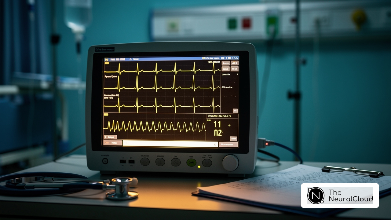
are characterized by their tall, peaked appearance on an ECG, frequently observed in the initial stages of myocardial infarction (MI). These patterns signify a critical alteration in the cardiac repolarization process, indicating stress or injury to the heart muscle. Clinically, recognizing hyperacute T waves can lead to timely interventions, potentially saving lives. The integration of technology enhances diagnostic accuracy. MaxYield™ employs advanced noise filtering and signal recognition, facilitating the rapid isolation and labeling of crucial ECG data, even amidst considerable noise and artifacts. Notably, MaxYield™ can analyze 200,000 heartbeats in under 5 minutes, enabling clinicians to swiftly identify abnormalities, which may suggest that the patient is experiencing acute coronary syndrome. This underscores the urgency for healthcare providers to act without delay. Understanding these signals not only aids in diagnosis but also enhances the overall patient care, emphasizing the importance of timely intervention in clinical practice.
Key Clinical Implications:
- Hyperacute T waves often serve as one of the earliest indicators of an impending heart attack, and MaxYield™ contributes to their precise identification. For instance, a 64-year-old man with a history of hypertension and diabetes presented with central chest discomfort radiating to his left arm, where an ECG revealed significant T changes, prompting urgent evaluation for myocardial infarction.
- Risk Stratification: Recognizing these patterns aids in assessing the severity of the cardiac event and guiding treatment decisions, supported by MaxYield™'s capabilities. In cases where individuals exhibit extreme T patterns, such as a 70-year-old man with acute chest discomfort, clinicians can prioritize interventions based on the urgency indicated by the ECG findings.
- Monitoring: Continuous ECG surveillance for early T signals provides real-time insights into a patient's cardiac status, enhanced by MaxYield™'s ability to manage large volumes of data. For example, a follow-up ECG conducted 90 minutes after the initial assessment displayed large, symmetric T deflections in a patient experiencing chest discomfort, highlighting the dynamic nature of cardiac events.
The importance of identifying rapid T phenomena, specifically hyperacute T waves, is emphasized by cardiologists, who assert that prompt recognition can significantly affect outcomes for patients. Recent studies have shown that sharp T patterns are vital not only for detecting myocardial infarction but also for differentiating it from other conditions, such as hyperkalemia and early repolarization patterns. This knowledge improves the overall diagnostic accuracy, reinforcing the necessity for vigilance in clinical practice.

Identify Key Characteristics and Diagnostic Criteria for Hyperacute T Waves
To accurately identify hyperacute T waves, clinicians should focus on several key characteristics:
- Height: Hyperacute T peaks typically exceed the height of the preceding R peak, often surpassing 5 mm.
- Peaked Appearance: These oscillations display a sharp, pointed apex, distinguishing them from the more rounded typical T waves.
- Symmetry: Hyperacute T forms are generally symmetrical, with both sides of the shape equal in height and duration.
- Lead Variability: They may manifest in multiple leads, particularly in the precordial leads (V2-V6), and are often more pronounced in leads that correspond to the affected area of the heart.
- Duration: The duration of hyperacute T waves is usually shorter than that of normal T waves, contributing to their unique appearance.
Diagnostic Criteria:
- Comparison with Previous ECGs: Comparison with previous ECGs is crucial for confirming the presence of hyperacute T waves. The ECG machine facilitates this by rapidly labeling P, QRS, and T onsets, allowing clinicians to easily compare current ECGs with previous ones for any significant changes.
- Clinical Context: Correlating ECG findings with the patient's clinical presentation is essential for accurate diagnosis. Regular serial 12-lead electrocardiograms should be acquired every 5 to 10 minutes to observe changes, as sharp T deflections can develop quickly. The interpretation of these analyses adapts to new data and improves diagnostic yield over time.
Specialists highlight that hyperacute T waves are very early T deflections and serve as the initial electrocardiographic indication of myocardial ischemia, emerging soon after coronary blockage and quickly progressing into ST-segment elevation. Joel T Levis, a Senior Emergency Physician, observes that 'the earliest-recognized electrocardiographic indication of myocardial ischemia is hyperacute T waves, which occur before ST-segment elevation.' This underscores the importance of early detection in clinical practice. Furthermore, it is essential to differentiate sharp T patterns from other conditions, such as hyperkalemia, to prevent misdiagnosis and guarantee suitable treatment. By utilizing technology through MaxYield™, clinicians can enhance their diagnostic capabilities and improve outcomes for those they treat.

Apply Knowledge of Hyperacute T Waves in Clinical Decision-Making
Once sharp T signals are recognized, practitioners must incorporate this information into their decision-making. Consider the following steps:
- Immediate Assessment: Evaluate the individual's symptoms, history, and risk factors for coronary artery disease. This context is crucial for determining the urgency of the situation.
- Further Testing: If sharp T forms are present, additional tests such as blood tests (e.g., troponin levels) and imaging studies may be necessary to confirm myocardial injury.
- Treatment Plan: Based on the findings, initiate appropriate interventions, which may include medications like antiplatelets, anticoagulants, or thrombolytics. Consider urgent referral for catheterization if indicated. Research shows that prompt action after the detection of rapid T patterns can greatly enhance outcomes for individuals, potentially decreasing the time to treatment by as much as 3 hours.
- Ongoing Surveillance: Establish ongoing monitoring to observe any alterations in the individual's cardiac condition, as elevated T patterns may progress into more serious irregularities. This is vital for intervention as needed.
- Documentation and Communication: Document findings and communicate with the healthcare team to ensure coordinated care and timely interventions. Effective communication is essential for optimizing patient management and ensuring that all team members are aware of the patient's evolving condition.
Utilizing Technology:
- Automation Tools: Leverage platforms like AI systems to automate the detection of abnormalities, enhancing diagnostic accuracy and efficiency in clinical workflows. AI technology transforms lengthy and noisy ECG recordings into clean, crisp signals, allowing healthcare professionals to make informed decisions based on reliable data. The integration of AI technology not only streamlines the identification process but also supports healthcare professionals in making informed decisions based on reliable data, as highlighted by the PMcardio platform's capabilities.
Identifying hyperacute T waves as crucial signs of myocardial infarction underscores the significance of prompt action. As noted by K. Thygesen, these waves can serve as STEMI equivalents, emphasizing the need for swift intervention to optimize patient outcomes.

Conclusion
Recognizing hyperacute T waves is essential for effective ECG interpretation, particularly in the context of acute myocardial infarction. These distinct, peaked waves serve as critical indicators of cardiac distress, enabling healthcare professionals to act swiftly and decisively. The integration of advanced tools like MaxYield™ enhances the ability to identify these patterns, ensuring timely diagnosis and intervention that can significantly impact patient outcomes.
The clinical implications of hyperacute T waves are profound, emphasizing their role in early detection of myocardial infarction, risk stratification, and ongoing patient monitoring. By understanding the key characteristics and diagnostic criteria, clinicians can better differentiate hyperacute T waves from other conditions, thereby improving the accuracy of their assessments. Immediate clinical decision-making following the identification of these waves is crucial, showcasing how rapid action can lead to better management of cardiac events.
Ultimately, mastering the interpretation of hyperacute T waves is vital in the realm of cardiology. As technology continues to evolve, leveraging AI-driven platforms like MaxYield™ will enhance diagnostic capabilities and facilitate better patient care. The urgency to recognize these early signs of cardiac distress is paramount; it is a vital skill that can save lives and improve the quality of care for patients experiencing acute coronary events.
Frequently Asked Questions
What are hyperacute T waves?
Hyperacute T waves are tall, peaked patterns observed on an ECG, often indicating significant alterations in cardiac repolarization, typically seen in the early stages of myocardial infarction (MI).
Why are hyperacute T waves clinically important?
They serve as early indicators of myocardial infarction, allowing for timely medical interventions that can potentially save lives.
How does MaxYield™ enhance the examination of hyperacute T waves?
MaxYield™ utilizes advanced noise filtering and signal recognition to quickly isolate and label crucial ECG data, enabling the analysis of 200,000 heartbeats in under 5 minutes, which helps clinicians swiftly identify hyperacute T waves.
What role do hyperacute T waves play in acute myocardial infarction?
They are often one of the earliest signs of an impending heart attack, prompting urgent evaluations and interventions.
How does recognizing hyperacute T waves assist in risk stratification?
Identifying these patterns helps assess the severity of a cardiac event and guides treatment decisions, allowing clinicians to prioritize interventions based on ECG findings.
What is the significance of continuous ECG surveillance for hyperacute T waves?
Continuous monitoring provides real-time insights into a patient's cardiac status, helping detect dynamic changes that may indicate worsening conditions.
How can hyperacute T waves help differentiate between different cardiac conditions?
Sharp T patterns are crucial for not only detecting myocardial infarction but also for distinguishing it from other issues like hyperkalemia and early repolarization patterns.
What is the overall impact of recognizing hyperacute T waves in clinical practice?
Prompt recognition of hyperacute T waves significantly affects patient outcomes and enhances the overall management of cardiac patients, highlighting the necessity for accurate ECG interpretation.
List of Sources
- Define Hyperacute T Waves and Their Clinical Importance
- pmc.ncbi.nlm.nih.gov (https://pmc.ncbi.nlm.nih.gov/articles/PMC4500486)
- thepermanentejournal.org (https://thepermanentejournal.org/doi/10.7812/TPP/14-243)
- powerfulmedical.com (https://powerfulmedical.com/blog/hyperacute-t-waves)
- ecgweekly.com (https://ecgweekly.com/ecg-tag/hyperacute-t-waves)
- emra.org (https://emra.org/emresident/article/stemi-equivalents)
- Identify Key Characteristics and Diagnostic Criteria for Hyperacute T Waves
- emra.org (https://emra.org/emresident/article/stemi-equivalents)
- pmc.ncbi.nlm.nih.gov (https://pmc.ncbi.nlm.nih.gov/articles/PMC4500486)
- powerfulmedical.com (https://powerfulmedical.com/blog/hyperacute-t-waves)
- Apply Knowledge of Hyperacute T Waves in Clinical Decision-Making
- annemergmed.com (https://annemergmed.com/article/S0196-0644(24)01250-2/fulltext)
- powerfulmedical.com (https://powerfulmedical.com/blog/hyperacute-t-waves)
- pubmed.ncbi.nlm.nih.gov (https://pubmed.ncbi.nlm.nih.gov/36774205)
- emra.org (https://emra.org/emresident/article/stemi-equivalents)
- ahajournals.org (https://ahajournals.org/doi/10.1161/CIR.0000000000001309)