Introduction
Understanding the complexities of an electrocardiogram (EKG) is crucial for anyone involved in cardiac health. It provides a glimpse into the heart's electrical activity, which is vital for assessing heart function. This article explores the essential components of a normal EKG, highlighting how these elements can reveal important information about heart health and potential abnormalities. Given the intricate nature of heart rhythms, healthcare professionals often face challenges in accurately distinguishing between normal and abnormal findings. This is where the MaxYield™ platform comes into play, enhancing ECG analysis and supporting accurate interpretations.
Define EKG: Understanding Its Role in Cardiac Health
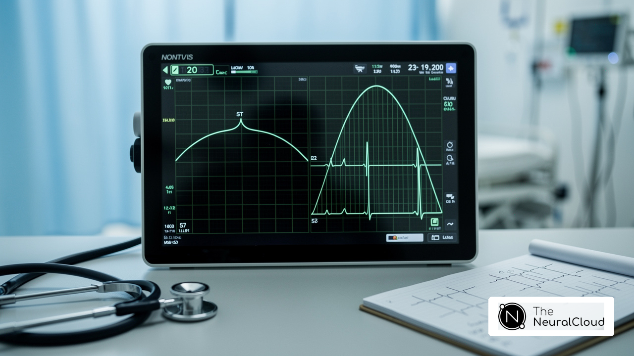
An electrocardiogram (EKG or ECG) is a diagnostic tool that records the electrical activity of the heart over time. It provides a visual representation of the heart's rhythm and electrical impulses, which can be compared to a standard reference to aid in the identification of various cardiac conditions. EKGs are crucial for diagnosing arrhythmias, heart attacks, and other heart-related issues. By analyzing the patterns and intervals of the EKG in comparison to a normal baseline, healthcare providers can assess the heart's condition and make informed treatment decisions.
Recent studies, including an analysis of over 208,000 ECGs from COPD cases, underscore the importance of EKGs in clinical settings, showcasing their effectiveness in identifying heart problems. For example, the use of EKG monitoring has been widely adopted and has successfully identified numerous instances of cardiac distress, significantly enhancing patient outcomes.
Cardiologists emphasize the value of EKGs in medical environments, noting that they provide essential insights into cardiac function. As Dr. Bryony Henderson stated, "GLP-1 agonists are pivotal in obesity care, promoting weight loss and addressing related health issues, with a focus on personalized, holistic treatment." Early detection of irregularities can be life-saving, making EKGs a vital component of modern cardiac care.
As the field evolves, the integration of AI-driven analysis, such as that offered by Neural Cloud Solutions' MaxYield™ platform, promises to refine EKG interpretation further. MaxYield™ enhances workflow efficiency through automation and advanced algorithms, allowing clinicians to transform noisy recordings into detailed insights. This ensures they can deliver the best care while effectively managing physiological variability and signal artifacts.
Features of MaxYield™
- Noise Reduction: Reduces background noise for clearer readings.
- Process Streamlining: Streamlines the interpretation process, saving time.
- Detailed Insights: Converts complex data into actionable information.
Advantages for Clinicians
- Reliability Enhancement: Enhances the reliability of EKG readings.
- Increased Efficiency: Allows for quicker decision-making in patient care.
- Better Patient Outcomes: Facilitates improved treatment strategies.

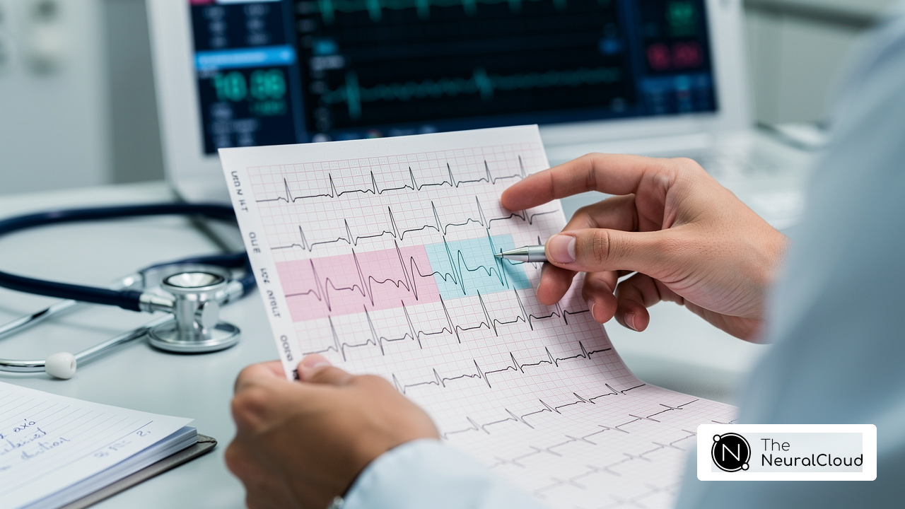
Analyze EKG Components: Intervals and Waveforms Explained
The EKG consists of several essential components, each reflecting distinct phases of the heart's electrical activity:
- P wave: This wave signifies atrial depolarization, marking the contraction of the atria. Irregularities in the P signal can suggest conditions like atrial fibrillation, which is vital for diagnosis.
- PR interval: Measuring the time from the onset of the P deflection to the beginning of the QRS complex, this interval typically ranges from 0.12 to 0.20 seconds. A prolonged PR interval may indicate atrioventricular block, necessitating further evaluation.
- QRS complex: Representing ventricular depolarization, the QRS complex indicates the electrical activity leading to ventricular contraction. A normal duration is less than 0.12 seconds; wider complexes may signal conduction abnormalities, such as bundle branch block.
- ST segment: This flat section between the QRS complex and the T crest reflects the period when the ventricles are fully depolarized. Subtle changes in the ST segment can indicate ischemia, making its assessment vital in clinical practice.
- T wave: The T component signifies ventricular repolarization, where the ventricles recover from contraction. Typically, T deflections are upright in most leads except aVR, where they may be inverted, emphasizing the significance of lead selection in interpretation.
- QT interval: Spanning from the start of the QRS complex to the conclusion of the T crest, this interval represents the total duration for ventricular depolarization and repolarization. A normal QT interval is less than or equal to 0.40 seconds; prolonged QT can increase the risk of arrhythmias, emphasizing the need for careful monitoring.
Understanding these components is crucial for healthcare professionals, as they help in comparing against a picture of normal EKG to gain insights into cardiac function and potential abnormalities. Recent findings underscore the importance of analyzing EKG waveforms and intervals, with ongoing research focusing on their clinical significance in diagnosing various cardiac conditions.

Prepare for EKG Testing: Steps and Expectations
To ensure a successful EKG test, patients should:
- Wear comfortable clothing: Opt for clothing that allows easy access to the chest, arms, and legs, making the process straightforward.
- Avoid Lotions and Creams: On the day of the test, skip any lotions and creams, as they can interfere with electrode adhesion and impact signal quality.
- Limit Stimulants: Steer clear of caffeine and alcohol before the test, since these substances can alter pulse rate and rhythm, potentially leading to misleading results.
- Inform the Technician: Share a complete list of medications and any existing medical conditions with the technician, as these factors can significantly affect EKG outcomes.
- Relax: Arrive at the testing facility 10-15 minutes early to unwind, as stress can affect heart rate.
- Follow Instructions: During the test, listen to the technician's guidance, including remaining still and breathing normally, to ensure accurate readings.
These steps not only enhance the reliability of EKG results but also align with the latest guidelines for EKG testing. By following these recommendations, patients can improve compliance rates and overall experiences during the testing process. Proper EKG preparation is crucial for accurate results, as inadequate preparation can lead to errors.

Interpret EKG Results: Identifying Normal and Abnormal Findings
Interpreting EKG results involves a comprehensive evaluation of the overall rhythm and the identification of any abnormalities. This process can be challenging due to the complexity of the data and the potential for misinterpretation. However, advancements in technology, such as artificial intelligence, are transforming ECG analysis, making it more accurate and efficient.
A normal EKG shows a consistent pattern of P waves, QRS complexes, and T waves, with a heart rate typically between 60 and 100 beats per minute. Recognizing a normal EKG and these normal patterns is crucial for healthcare professionals to identify deviations that may indicate underlying conditions.
Abnormal Findings: Deviations from the normal EKG pattern can signal various conditions:
- Arrhythmias: irregular heart rhythms can be detected.
- Myocardial Infarction: findings include ST segment elevation or depression. Recent studies highlight that technology, like that offered by AI platforms, can enhance detection while reducing unnecessary emergency activations. For example, an AI model identified 553 confirmed STEMIs compared to 427 detected by standard triage, showcasing the platform's advanced noise filtering and wave recognition capabilities.
- Prolonged QT Interval: This finding increases the risk of life-threatening arrhythmias, making timely intervention essential.
Clinical Correlation: Correlating EKG findings with clinical symptoms and patient history is vital for accurate diagnosis and effective management. As noted by Mumma, "STEMI is a life-threatening emergency where every minute matters," underscoring the urgency of recognizing these abnormalities. Leveraging a continuous learning model, healthcare professionals can improve the accuracy and efficiency of ECG analysis, addressing challenges such as physiological variability and signal artifacts. This not only enhances patient outcomes but also streamlines the workflow in emergency situations.

Conclusion
Understanding EKGs is crucial for healthcare professionals and patients alike. This article has highlighted the essential role electrocardiograms play in monitoring cardiac health. These diagnostic tools capture the heart's electrical activity, aiding in the identification of various conditions. By comparing EKG results to a standard EKG, clinicians can make informed decisions that significantly enhance patient care.
Key components of the EKG include:
- P wave
- QRS complex
- T wave
These components were examined in detail, showcasing their individual importance in diagnosing heart conditions. The article also stressed the necessity of proper preparation for EKG testing, which ensures reliable results that can lead to timely interventions. Furthermore, advancements in technology, including AI-driven analysis, promise to improve the accuracy and efficiency of EKG interpretation, ultimately benefiting patient outcomes.
The importance of understanding EKGs cannot be overstated. As cardiac health remains a pressing concern, gaining knowledge about EKG components and their implications fosters better health decisions. Whether preparing for an EKG test or interpreting results, being informed empowers individuals to take charge of their heart health and engage in proactive discussions with healthcare providers.
Frequently Asked Questions
What is an EKG and what does it measure?
An electrocardiogram (EKG or ECG) is a diagnostic tool that records the electrical activity of the heart over time, providing a visual representation of the heart's rhythm and electrical impulses.
How are EKGs used in diagnosing heart conditions?
EKGs are crucial for diagnosing arrhythmias, myocardial infarctions, and other heart-related issues by analyzing the patterns and intervals in comparison to a normal EKG.
What recent studies highlight the importance of EKGs?
Recent studies, including an analysis of over 208,000 ECGs from COPD cases, demonstrate the effectiveness of EKGs in detecting critical cardiac events.
What is the Kardia 12L system and its significance?
The Kardia 12L system is a widely adopted tool that has successfully identified numerous instances of myocardial infarction, significantly improving patient outcomes.
Why do cardiologists emphasize the value of EKGs?
Cardiologists highlight EKGs for providing essential insights into cardiac function, which can lead to early detection of irregularities that may be life-saving.
How is AI being integrated into EKG analysis?
AI-driven analysis, such as Neural Cloud Solutions' MaxYield™ platform, enhances EKG interpretation through advanced noise filtering and automated analysis, improving the quality of readings.
What are the features of the MaxYield™ platform?
The MaxYield™ platform includes advanced noise filtering, automated analysis, and the ability to convert complex data into actionable insights.
What advantages does MaxYield™ offer to healthcare professionals?
MaxYield™ improves accuracy of EKG readings, increases efficiency in patient care, and facilitates better patient outcomes through timely interventions based on precise data.
List of Sources
- Define EKG: Understanding Its Role in Cardiac Health
- HeartBeam Receives FDA Clearance for First-Ever, Cable-Free Synthesized 12-Lead ECG for At-Home Arrhythmia Assessment (https://ir.heartbeam.com/news-events/press-releases/detail/107/heartbeam-receives-fda-clearance-for-first-ever-cable-free)
- news-medical.net (https://news-medical.net/news/20251103/More-frequent-use-of-electrocardiograms-could-help-prevent-sudden-cardiac-death.aspx)
- AI-Powered ECG Analysis Offers Promising Path for Early Detection of Chronic Obstructive Pulmonary Disease, Say Mount Sinai Researchers (https://mountsinai.org/about/newsroom/2026/ai-powered-ecg-analysis-offers-promising-path-for-early-detection-of-chronic-obstructive-pulmonary-disease-say-mount-sinai-researchers)
- medicaldevice-network.com (https://medicaldevice-network.com/news/alivecor-fda-nod-kardia-system)
- biospace.com (https://biospace.com/press-releases/alivecor-announces-fda-clearance-of-new-cardiac-determinations-for-kardia-12l-ecg-system-bringing-total-to-39)
- Analyze EKG Components: Intervals and Waveforms Explained
- AI-powered ECG model outperforms doctors in detecting hidden heart disease (https://news-medical.net/news/20250721/AI-powered-ECG-model-outperforms-doctors-in-detecting-hidden-heart-disease.aspx)
- geekymedics.com (https://geekymedics.com/ecg-case-studies)
- ECG Learning Center - An introduction to clinical electrocardiography (https://ecg.utah.edu/lesson/3)
- bookey.app (https://bookey.app/book/the-ecg-made-easy/quote)
- Normal Electrocardiography (ECG) Intervals: Normal Electrocardiography Intervals (https://emedicine.medscape.com/article/2172196-overview)
- Prepare for EKG Testing: Steps and Expectations
- int.livhospital.com (https://int.livhospital.com/ekg-best-preparation-tips-for-success)
- Accuracy of Physicians’ Electrocardiogram Interpretation (https://jamanetwork.com/journals/jamainternalmedicine/fullarticle/2771093)
- jamanetwork.com (https://jamanetwork.com/journals/jamainternalmedicine/fullarticle/2820721)
- Electrocardiogram - StatPearls - NCBI Bookshelf (https://ncbi.nlm.nih.gov/books/NBK549803)
- danleemedical.com (https://danleemedical.com/blog/everything-you-need-to-know-about-ekg-prep)
- Interpret EKG Results: Identifying Normal and Abnormal Findings
- news-medical.net (https://news-medical.net/news/20251029/AI-driven-ECG-improves-detection-of-severe-heart-attacks.aspx)
- New study finds AI model improves heart attack detection (https://health.ucdavis.edu/news/headlines/new-study-finds-ai-model-improves-heart-attack-detection/2025/11)
- cureus.com (https://cureus.com/articles/289924-incidence-and-prognostic-significance-of-arrhythmia-in-acute-myocardial-infarction-presentation-an-observational-study)
- thecardiologyadvisor.com (https://thecardiologyadvisor.com/news/routine-ecg-screening-may-prevent-cardiovascular-disease-events)
- tctmd.com (https://tctmd.com/news/ecg-abnormalities-routine-screening-tied-cvd-events-large-study)