Overview
The article provides an overview of the challenges associated with diagnosing anterior wall myocardial infarction (AWMI) through ECG. It emphasizes the importance of recognizing specific ECG changes, such as ST-segment elevation and Q waves, which are critical for accurate diagnosis. Timely recognition and analysis of these changes are essential for improving patient outcomes. The use of advanced systems like MaxYield™ enhances ECG analysis, enabling healthcare professionals to respond promptly with appropriate treatment interventions.
MaxYield™ offers several features that significantly improve ECG analysis. By facilitating the identification of crucial ECG changes, the platform streamlines the diagnostic process. This results in a more efficient workflow for healthcare professionals, allowing them to focus on patient care. The advantages of using MaxYield™ extend beyond mere diagnosis; they include enhanced accuracy and faster decision-making capabilities, ultimately leading to better patient outcomes.
Incorporating visual aids, such as diagrams or infographics, can further clarify complex information presented in the article. This approach not only enhances understanding but also engages the reader, making the content more accessible. By maintaining a balance between informative sections and succinct summaries, the article ensures that readers remain engaged while comprehending the critical information.
Overall, the article highlights the pivotal role of timely ECG analysis in diagnosing AWMI and underscores how the MaxYield™ platform can transform this process for healthcare professionals, leading to improved patient care.
Introduction
Understanding the nuances of anterior wall myocardial infarction is essential for healthcare professionals, as timely diagnosis can significantly influence patient outcomes. This guide explores the critical ECG changes associated with this condition, highlighting how advanced tools like the MaxYield™ system can enhance diagnostic accuracy and efficiency. As the complexities of ECG interpretation grow, clinicians must not only identify these crucial changes but also effectively address the common challenges that arise during analysis.
Understand Anterior Wall Myocardial Infarction
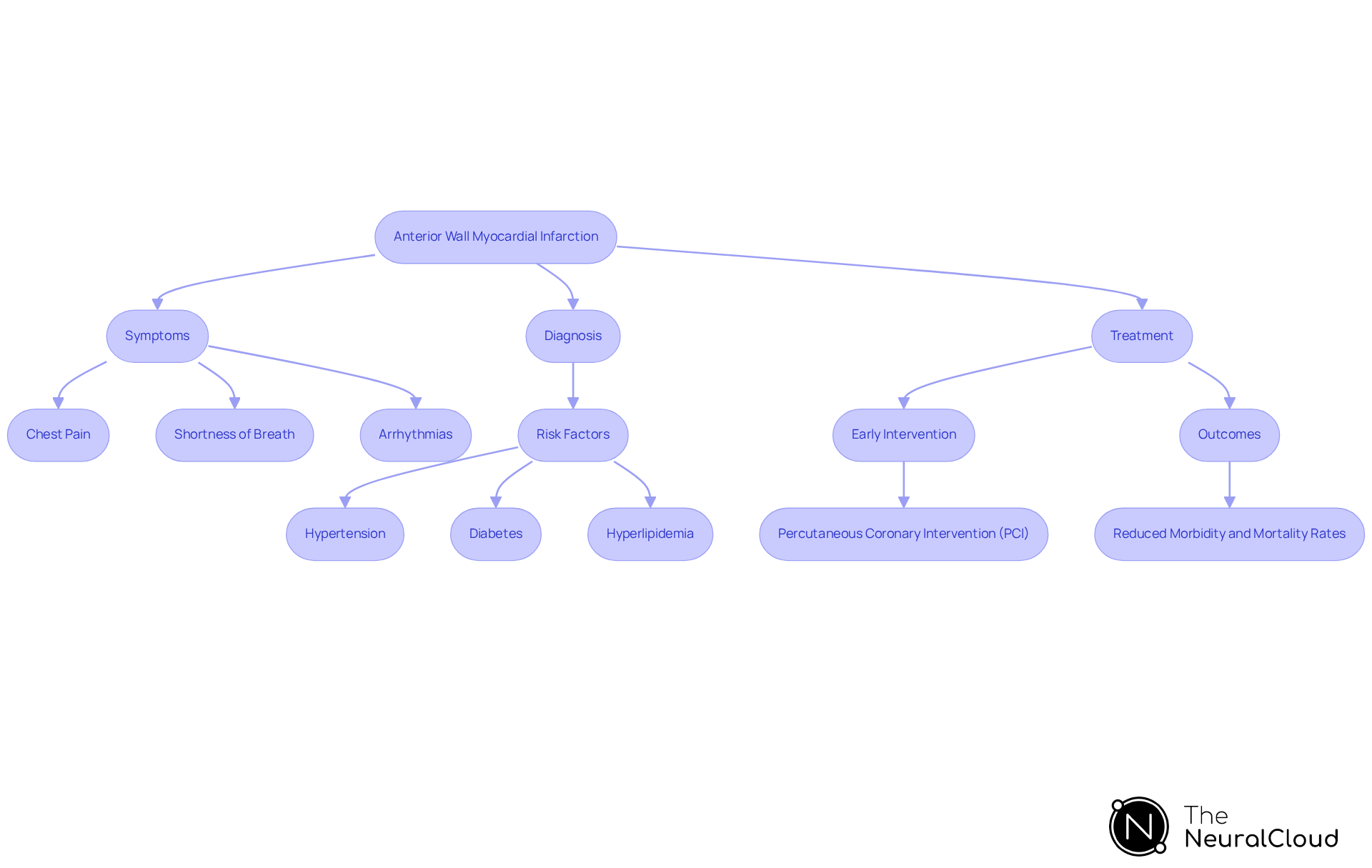
Anterior wall myocardial infarction happens when there is an obstruction of blood flow to the heart's front wall, usually caused by a blockage in the left anterior descending artery. This condition can lead to significant cardiac damage if not diagnosed and treated promptly. Key symptoms include chest pain, shortness of breath, and potential arrhythmias, which necessitate immediate medical attention. Understanding the fundamental mechanisms of is crucial for healthcare professionals, as it informs the urgency of intervention and directs treatment strategies.
Risk factors for acute myocardial infarction include hypertension, diabetes, and hyperlipidemia, which are common in many patients. For instance, nearly 47% of U.S. adults are affected by high blood pressure, and over 72% have an unhealthy weight, both of which significantly increase the risk of cardiovascular events. Additionally, the incidence of heart failure (HF) after anterior wall myocardial infarction (MI) varies widely, with studies indicating a 1-year incidence ranging from 2% to 30%, depending on the definition criteria used.
Prompt diagnosis plays a crucial role in enhancing outcomes for patients with acute wall motion impairment. Research shows that early intervention, such as percutaneous coronary intervention (PCI), can significantly reduce morbidity and mortality rates. Cardiologists stress that identifying the symptoms and risk factors related to acute wall motion abnormalities can result in faster treatment choices, ultimately maintaining heart function and improving patient survival rates. As advancements in ECG technology continue to evolve, healthcare professionals must remain vigilant in identifying and managing this life-threatening condition.

Identify Key ECG Changes in Anterior Wall Infarction
In diagnosing anterior wall myocardial infarction, specific ECG changes serve as essential indicators of the condition. Key features to identify include:
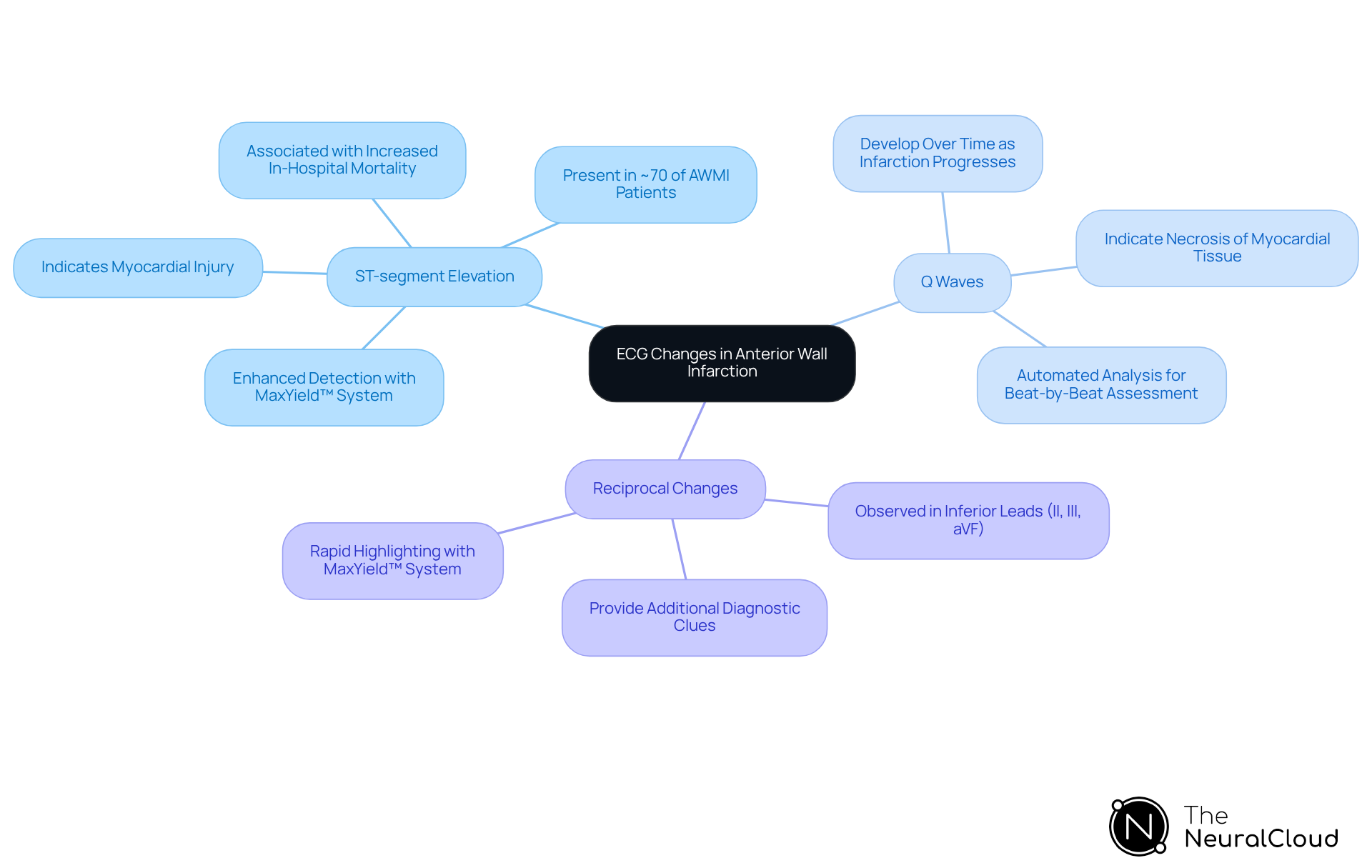
- ST-segment elevation in leads V1 to V4, signifying myocardial injury and present in approximately 70% of patients with AWMI. This elevation is critical because anterior wall myocardial infarction (AWMI) is associated with increased in-hospital mortality and a greater reduction in left ventricle ejection fraction compared to other types of myocardial infarction. The MaxYield™ system enhances the detection of this elevation by mapping ECG signals through noise, ensuring that healthcare professionals can quickly identify these critical changes.
- Q waves in these leads indicate necrosis of myocardial tissue, which can develop over time as the infarction progresses. The system provides automated analysis capabilities that allow for beat-by-beat assessments, offering detailed insights into the progression of these changes, including the identification of Q waves.
- Reciprocal changes may also be observed in inferior leads (II, III, aVF), providing additional diagnostic clues that can aid in confirming the diagnosis. With this system, these reciprocal changes can be , allowing for a more comprehensive understanding of the patient's condition.
Healthcare professionals must recognize these changes promptly, as they are vital for diagnosing anterior wall myocardial infarction and initiating timely treatment. The incorporation of the MaxYield™ system into clinical workflows not only aids in recognizing these ECG patterns but also significantly influences patient outcomes, especially in emergency environments where swift intervention is crucial. Furthermore, the Fourth Universal Definition of Myocardial Infarction established in 2018 underscores the importance of these diagnostic criteria, while elevated cardiac biomarkers also play a critical role in confirming myocardial infarction.

Analyze ECG Data Using MaxYield™ for Accurate Diagnosis
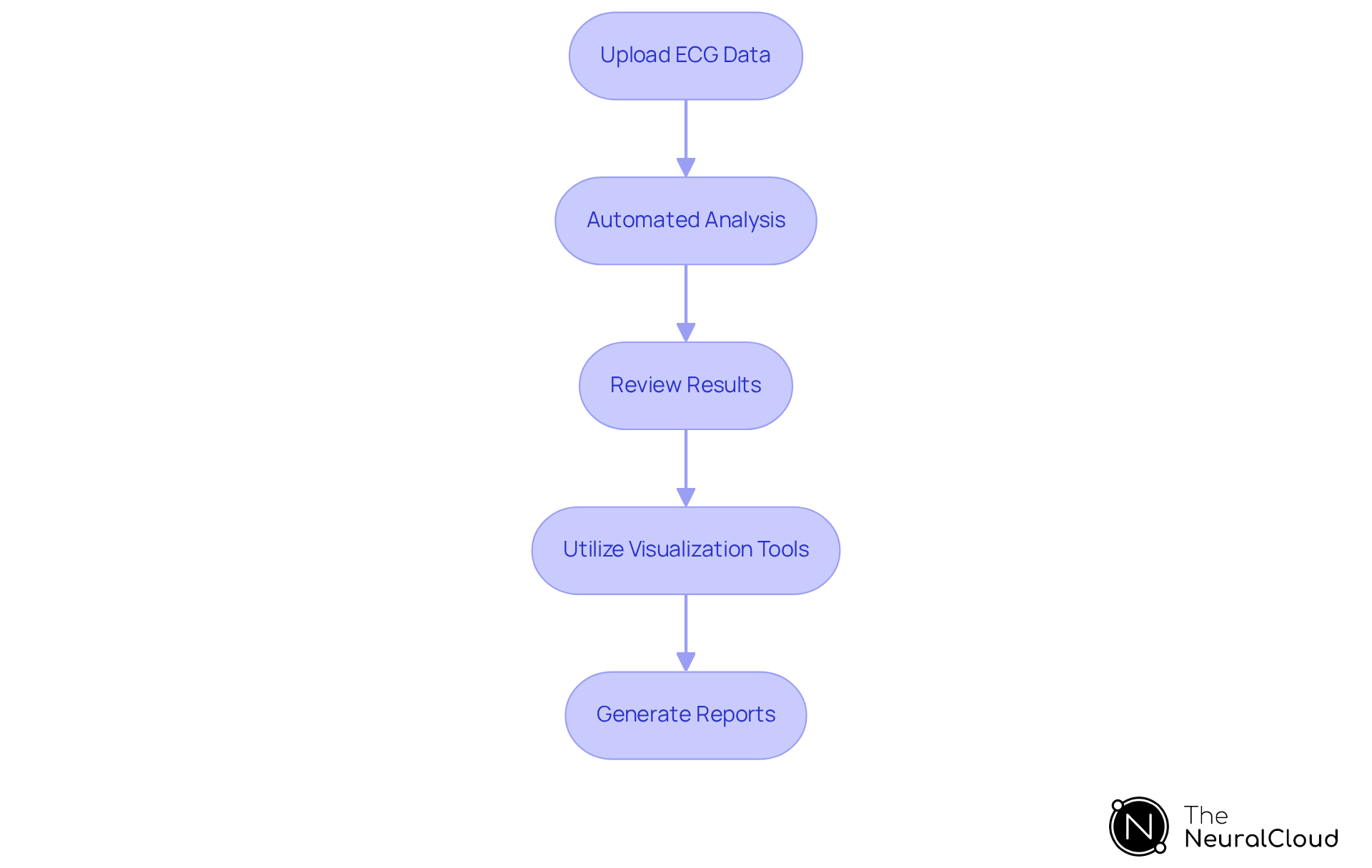
To effectively analyze ECG data using the MaxYield™ platform, clinicians can follow these streamlined steps:
- Upload ECG Data: Import ECG recordings from various devices, including Holter monitors and wearable devices, into the MaxYield™ system.
- Automated Analysis: The system processes the data, automatically labeling key features such as P-waves, QRS complexes, and T-wave intervals. This automation enhances both the speed and accuracy of the analysis.
- Review Results: Examine the output for critical indicators like ST-segment elevations and Q waves in leads V1-V4, which are essential for diagnosing anterior wall myocardial infarction (AWMI).
- Utilize Visualization Tools: Leverage the system's to clarify ECG changes. This facilitates effective communication of findings with colleagues.
- Generate Reports: Create comprehensive reports summarizing the analysis. These reports can be shared with the clinical team for informed decision-making.
By employing this advanced system, clinicians can save over 25% in analysis time compared to conventional methods, processing more than 200,000 heartbeats in under five minutes. This efficiency not only reduces the workload associated with manual ECG interpretation but also enhances diagnostic accuracy. Evidence shows a 14-fold reduction in missed diagnoses of severe arrhythmias. Testimonials from healthcare professionals highlight the system's transformative effect on clinical workflows, emphasizing its contribution to improving patient outcomes through timely and accurate ECG analysis. Continuous advancements in automated ECG analysis technology ensure that clinicians have access to the latest capabilities, reinforcing its role as a vital tool in modern cardiac care.

Troubleshoot Common ECG Interpretation Challenges
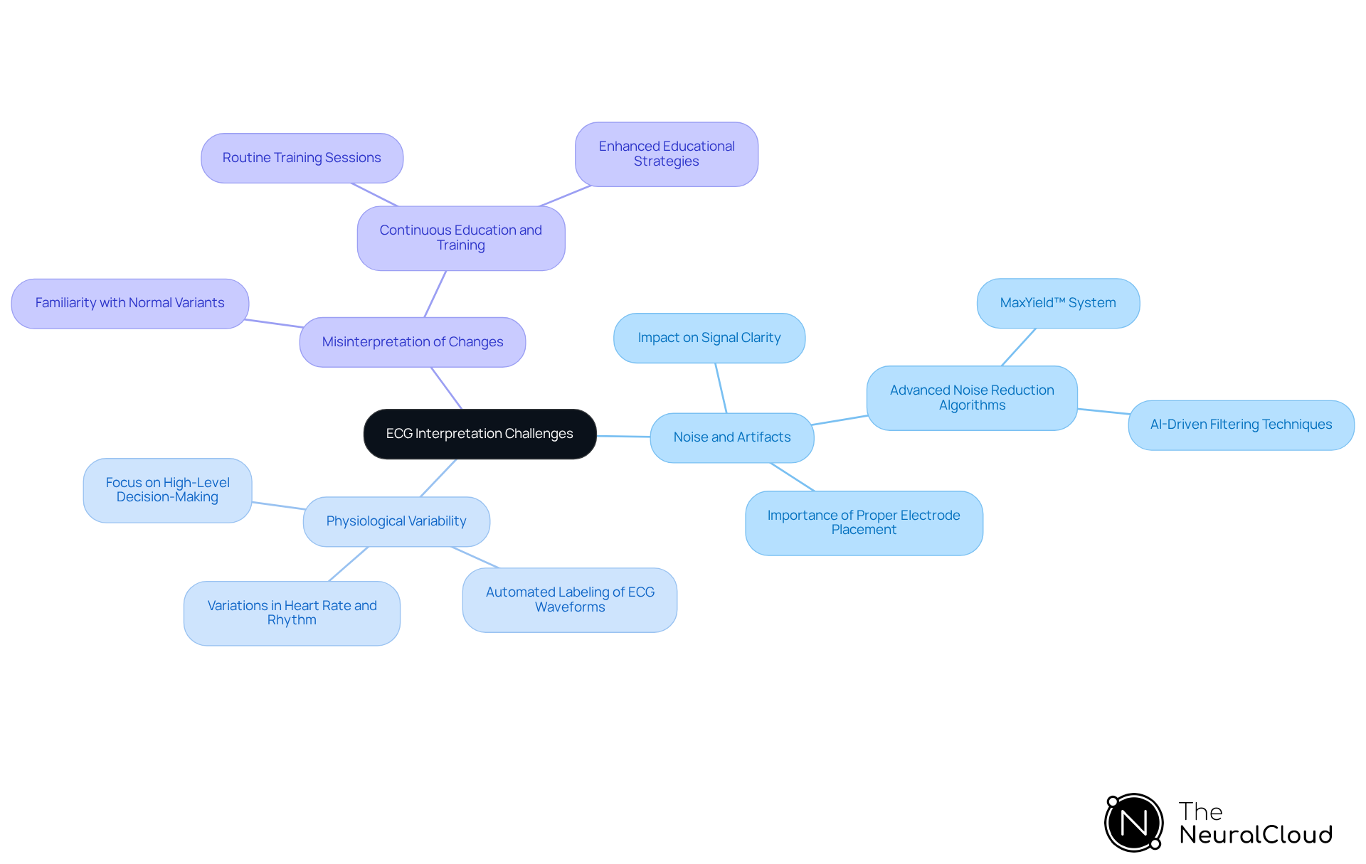
Common challenges in ECG interpretation include:
- Noise and Artifacts: A significant percentage of ECG readings are affected by noise and artifacts, obscuring true cardiac signals. Advanced noise reduction algorithms, such as those employed in the MaxYield™ system, are crucial for enhancing signal clarity and optimizing workflow through gold standard techniques. When noise is present, re-evaluating electrode placement or utilizing the system's filtering options can substantially improve data quality. Cardiologists emphasize the importance of effective noise management for accurate diagnoses, as even minor artifacts can lead to misinterpretation of critical cardiac events. As noted by Shifa Kaushal, 'ECG needs an effective denoising technique to ensure the integrity of the data.'
- Physiological Variability: Variations in heart rate and rhythm can complicate ECG interpretation. The MaxYield™ system is designed to account for these physiological changes, enabling more precise analysis. For instance, the system's automated labeling of ECG waveforms helps reduce the impact of variability, allowing healthcare professionals to focus on high-level decision-making rather than manual adjustments. This automation not only enhances accuracy but also increases the efficiency of ECG analysis.
- Misinterpretation of Changes: Familiarity with normal variants in ECG findings is crucial to avoid misdiagnosis. Continuous education and practice are essential components in improving interpretation skills. Routine training sessions, supported by the advanced features of the MaxYield™ system, can assist clinicians in staying informed on best practices and boosting their confidence in ECG analysis. The need for across the practice continuum is vital, as highlighted in recent studies.
For further assistance, consult the MaxYield™ user manual, which provides detailed guidance on effectively utilizing the platform, or reach out to technical support. Engaging in regular training can also strengthen interpretation skills and ensure accurate assessments.

Conclusion
Understanding and diagnosing anterior wall myocardial infarction (AWMI) is critical for effective patient care. This guide emphasizes the importance of recognizing the specific ECG changes associated with this condition, which are vital for timely intervention. By leveraging advanced tools like the MaxYield™ system, healthcare professionals can enhance diagnostic accuracy and significantly improve patient outcomes.
Key insights discussed include:
- The identification of ST-segment elevations and Q waves in specific leads, which serve as crucial indicators of myocardial injury and necrosis.
- The significance of addressing common challenges in ECG interpretation, such as noise and physiological variability, which can obscure critical cardiac signals.
By utilizing advanced technology and continuous education, clinicians can navigate these challenges and ensure accurate assessments.
Ultimately, the ability to swiftly diagnose anterior wall myocardial infarction through precise ECG interpretation not only saves lives but also preserves heart function. As advancements in ECG technology continue to evolve, embracing these tools and strategies will be essential for healthcare professionals dedicated to delivering high-quality cardiac care. Empowering clinicians with the knowledge and resources to excel in ECG interpretation is a crucial step toward enhancing patient survival rates and overall health outcomes in the face of myocardial infarction.
Frequently Asked Questions
What is anterior wall myocardial infarction?
Anterior wall myocardial infarction occurs when there is an obstruction of blood flow to the heart's front wall, typically due to a blockage in the left anterior descending artery.
What are the key symptoms of anterior wall myocardial infarction?
Key symptoms include chest pain, shortness of breath, and potential arrhythmias, which require immediate medical attention.
What are the major risk factors for acute myocardial infarction?
Major risk factors include hypertension, diabetes, and hyperlipidemia. For example, nearly 47% of U.S. adults have high blood pressure, and over 72% have an unhealthy weight, both of which increase the risk of cardiovascular events.
What is the incidence of heart failure after anterior wall myocardial infarction?
The incidence of heart failure after anterior wall myocardial infarction varies widely, with studies showing a 1-year incidence ranging from 2% to 30%, depending on the definition criteria used.
Why is prompt diagnosis important for anterior wall myocardial infarction?
Prompt diagnosis is crucial as it enhances patient outcomes. Early intervention, such as percutaneous coronary intervention (PCI), can significantly reduce morbidity and mortality rates.
How can healthcare professionals improve treatment outcomes for patients with anterior wall myocardial infarction?
By identifying symptoms and risk factors related to acute wall motion abnormalities, healthcare professionals can make faster treatment choices that help maintain heart function and improve patient survival rates.
What advancements are impacting the management of anterior wall myocardial infarction?
Advancements in ECG technology are evolving, allowing healthcare professionals to better identify and manage this life-threatening condition.


