Overview
The article highlights the clinical significance and identification of DeWinter T-waves in ECG analysis. These T-waves indicate acute occlusion in the left anterior descending artery and can signal the evolution of ST-Elevation Myocardial Infarction (STEMI). Recognizing these T-waves is crucial for timely intervention, as they may appear without the typical ST-segment elevation.
To address the challenges in ECG analysis, the article discusses the MaxYield™ platform, which enhances the detection of these critical T-waves. The platform utilizes advanced technology to improve the accuracy of ECG readings, ensuring that healthcare professionals can identify potential STEMI cases more effectively.
The features of the MaxYield™ platform include its ability to analyze complex ECG data quickly and accurately. This efficiency allows for prompt decision-making in clinical settings, ultimately benefiting patient outcomes. By integrating this technology into their practice, healthcare professionals can enhance their diagnostic capabilities and provide better care.
In summary, the MaxYield™ platform not only streamlines ECG analysis but also empowers healthcare providers with the tools necessary for timely interventions. The ability to detect DeWinter T-waves accurately can significantly impact patient management and treatment strategies.
Introduction
Recognizing subtle yet critical patterns in ECG readings can be the difference between life and death in acute cardiac situations. Among these patterns, DeWinter T-waves stand out as a significant indicator of evolving ST-Elevation Myocardial Infarction (STEMI), often linked to left anterior descending artery occlusion. This article delves into the essential characteristics, identification techniques, and clinical implications of DeWinter T-waves. Mastering this knowledge can empower healthcare professionals to make timely and potentially lifesaving decisions. However, how can clinicians ensure they do not overlook these crucial signs that may appear without the usual markers of cardiac distress?
Define DeWinter T-Waves: Key Characteristics and Clinical Relevance
This pattern is characterized by an upward-sloping ST-segment depression followed by prominent, symmetrical waves, typically observed in the precordial leads (V1-V6). It is often linked to acute occlusion of the left anterior descending artery (LAD) and serves as a crucial indicator of evolving ST-Elevation Myocardial Infarction (STEMI). Clinically, the recognition of these waves is essential, as they may appear without the conventional ST-segment elevation, presenting a concealed yet significant sign of cardiac ischemia.
The MaxYield™ platform from Neural Cloud Solutions enhances the detection of such critical patterns by utilizing advanced noise filtering and automated ECG analysis. This technology facilitates the rapid identification of T-waves even in recordings affected by high levels of noise and artifact. MaxYield™ provides beat-by-beat analysis and delivers detailed metrics, including P-wave, QRS complex, and T-wave onsets and offsets, which are vital for accurate diagnosis.
This specific pattern, known as deWinter's T-waves and originally outlined by Dr. DeWinter in 2008, is believed to occur in approximately 2% of individuals with anterior myocardial infarction. By integrating MaxYield™ into clinical workflows, health tech developers can ensure that clinicians are equipped with the necessary tools for swift recognition and intervention, potentially improving outcomes through timely action.
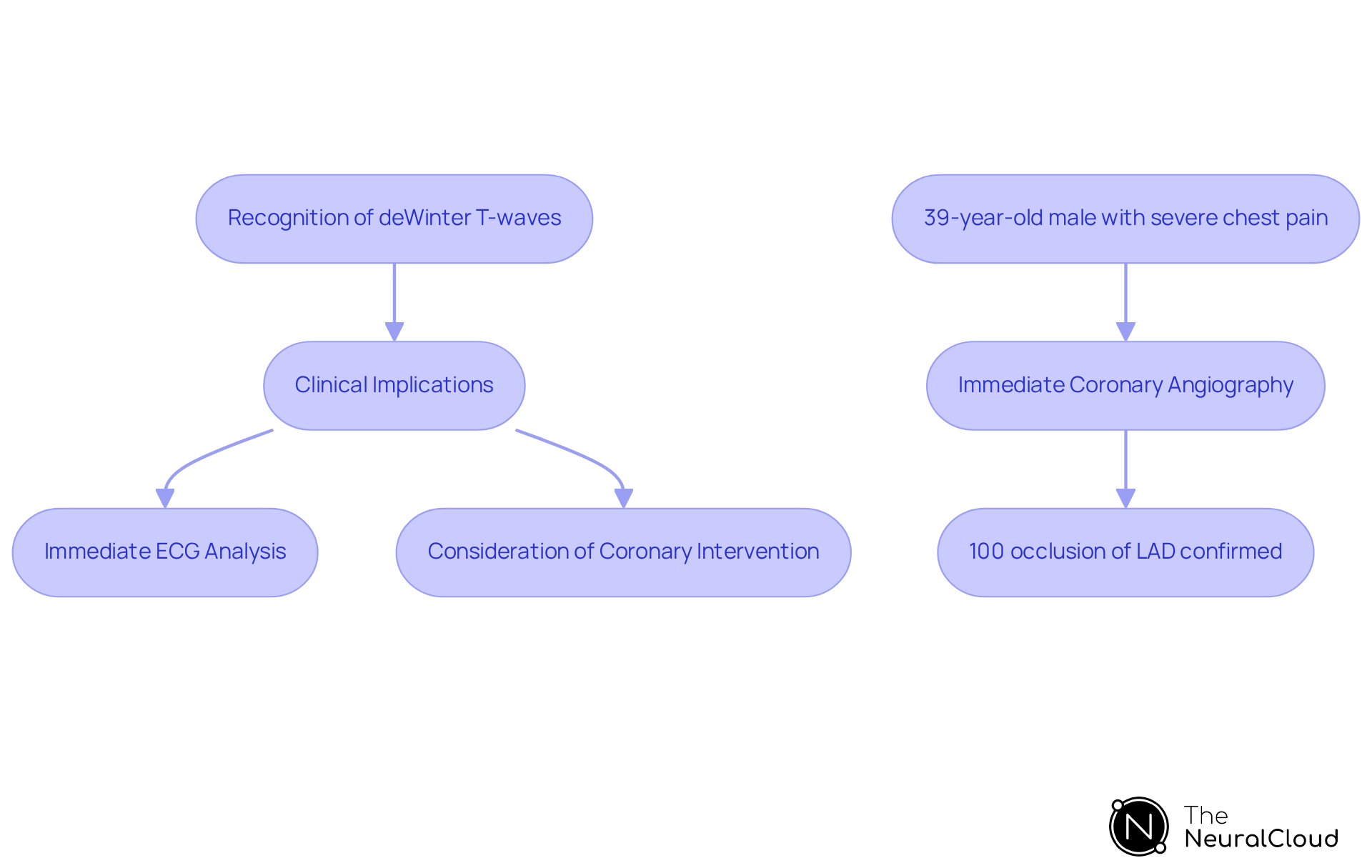
For example, in a notable case, a 39-year-old male presented with severe chest pain and diaphoresis, exhibiting pulseless polymorphic ventricular tachycardia during transport. His initial ECG displayed the specific pattern, prompting immediate coronary angiography, which confirmed a 100% thrombotic occlusion of the LAD. This case highlights the importance of heightened awareness regarding atypical ECG findings, as timely recognition can enable prompt coronary intervention and significantly enhance patient prognosis.

Identify DeWinter T-Waves: ECG Interpretation Techniques
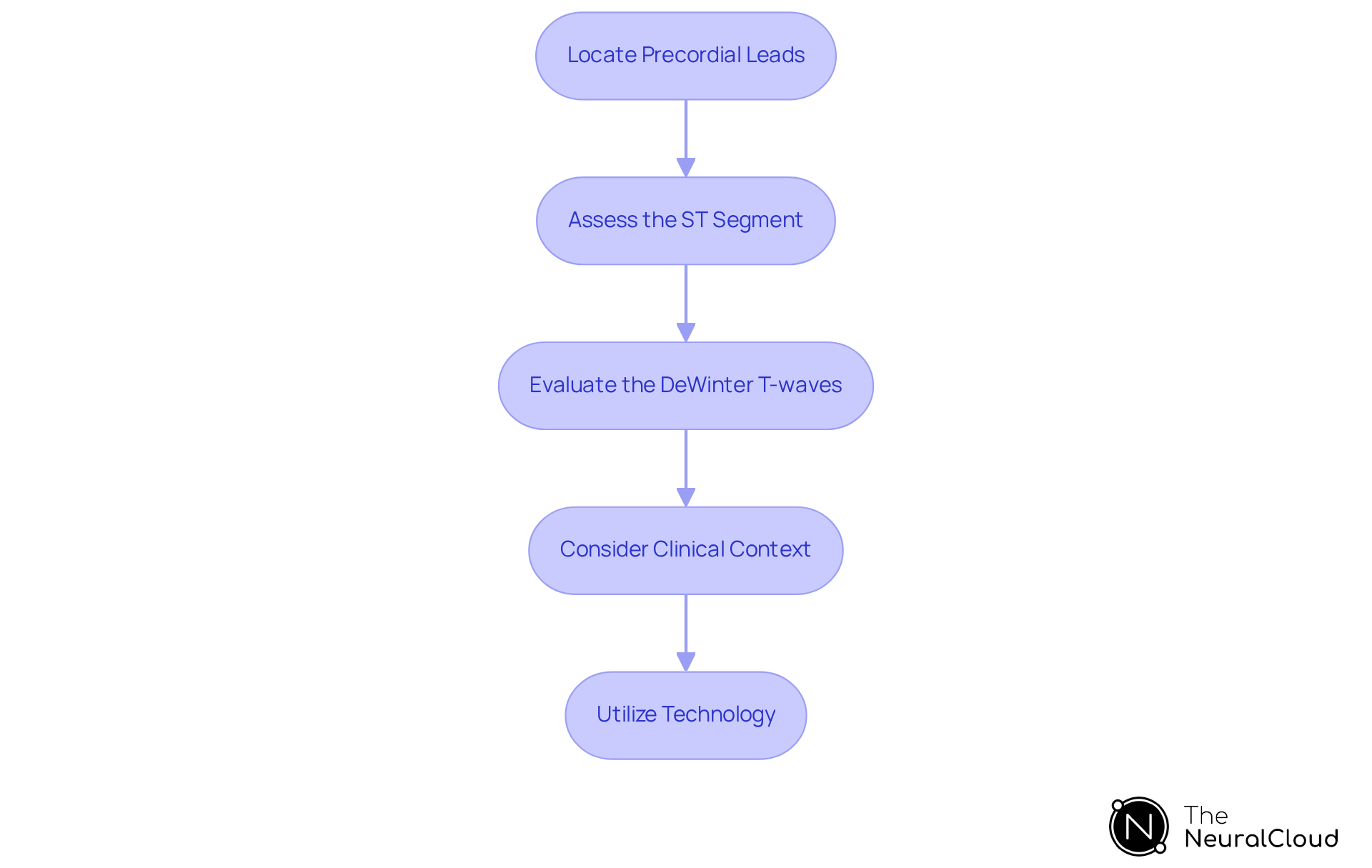
To accurately identify DeWinter T-waves on an ECG, follow these essential steps:
- Locate the Precordial Leads: Focus on leads V1 to V6, where the deWinter T-waves pattern is predominantly observed.
- Assess the ST Segment: Look for upsloping ST-segment depression at the J-point, typically ranging from 1 to 3 mm. This depression should exhibit a concave upward shape.
- Evaluate the DeWinter T-waves: Identify tall, symmetrical T-morphologies that follow the ST-segment depression. These transmembrane waves should be prominent and upright, indicating significant cardiac events.
- Consider Clinical Context: Correlate the ECG findings with the individual's clinical presentation, particularly symptoms of chest pain or indicators of acute coronary syndrome.
- Utilize Technology: Employ advanced ECG analysis tools, such as Neural Cloud Solutions' MaxYield™ platform, which automates the identification and labeling of specific wave patterns, thereby enhancing diagnostic efficiency.
By mastering these techniques, clinicians can significantly improve their ability to recognize this critical ECG finding, facilitating timely and appropriate management of patients with potential cardiac issues. The significance of identifying specific patterns is underscored by case studies where prompt recognition resulted in successful interventions, highlighting the necessity for heightened awareness within the healthcare field.

Analyze Clinical Implications: Diagnosing and Managing Conditions Associated with DeWinter T-Waves
The occurrence of T-waves on an ECG is a crucial sign of acute coronary syndrome, particularly in cases of proximal left anterior descending (LAD) artery blockage. Clinicians should be aware of the following clinical implications:
-
Immediate Action Required: The specific pattern necessitates urgent evaluation and intervention, often requiring the activation of the cardiac catheterization lab for potential percutaneous coronary intervention (PCI). Research indicates that this pattern is noted in approximately 2% of individuals with acute LAD occlusions, with the left anterior descending artery involved in 73% of instances, underscoring the necessity for swift action.
-
Differential Diagnosis: While specific pattern waves are indicative of severe coronary artery disease, they must be distinguished from other conditions such as Wellens syndrome or hyperkalemia, which can present with similar ECG changes. Misinterpretation can lead to delays in treatment, negatively impacting outcomes for individuals.
-
Patient Management: Patients exhibiting dewinters t waves should be closely monitored for signs of myocardial infarction and managed according to STEMI protocols. This involves providing antiplatelet treatment and evaluating thrombolysis, as prompt intervention can significantly enhance reperfusion rates. Research shows nearly 80% success in individuals receiving systemic thrombolysis, with 82% demonstrating clinical improvement.
-
Long-term Considerations: Following acute management, individuals may require additional assessment for underlying coronary artery disease. This may involve stress testing or coronary angiography to evaluate the necessity for ongoing treatment or intervention, particularly since 93% of individuals with the syndrome experience severe to total occlusion of at least one coronary vessel. Moreover, it is crucial to recognize that there is a reported 3% mortality rate during the same admission for individuals presenting with dewinters t waves, along with a 9% incidence rate of malignant ventricular arrhythmia linked to this syndrome.
By understanding these clinical implications, healthcare professionals can enhance their decision-making processes, ultimately improving patient outcomes in cases of acute coronary syndrome. As emphasized by experts, 'Early diagnosis of the dewinters t waves ECG pattern can be lifesaving,' highlighting the critical nature of recognizing this pattern in clinical practice.

Conclusion
Mastering the identification of DeWinter T-waves is crucial for healthcare professionals engaged in ECG analysis. Recognizing these specific patterns not only aids in diagnosing acute coronary syndromes but also plays a vital role in determining timely interventions that can significantly impact patient outcomes. The integration of advanced technologies, such as the MaxYield™ platform, enhances the ability to detect these critical signs amidst challenging conditions.
Key insights from this article highlight the characteristics of DeWinter T-waves, their association with severe cardiac events, and the imperative for immediate action upon their identification. By understanding the nuances of ECG interpretation techniques, clinicians can effectively differentiate DeWinter T-waves from other similar patterns, ensuring that patients receive the appropriate management and care. The importance of swift clinical response cannot be overstated, as timely recognition can lead to life-saving interventions.
Ultimately, the significance of DeWinter T-waves extends beyond mere identification; it encompasses a broader commitment to improving clinical practices and patient safety. As the landscape of cardiology continues to evolve, staying informed about current research and advancements in ECG analysis will empower healthcare professionals to enhance their diagnostic capabilities. This proactive approach is essential for fostering better health outcomes and underscores the critical nature of recognizing DeWinter T-waves in clinical settings.
Frequently Asked Questions
What are DeWinter T-waves and how are they characterized?
DeWinter T-waves are characterized by an upward-sloping ST-segment depression followed by prominent, symmetrical waves, typically observed in the precordial leads (V1-V6).
What is the clinical significance of DeWinter T-waves?
DeWinter T-waves are often linked to acute occlusion of the left anterior descending artery (LAD) and serve as a crucial indicator of evolving ST-Elevation Myocardial Infarction (STEMI). Their recognition is essential, as they may appear without the conventional ST-segment elevation, indicating significant cardiac ischemia.
How does the MaxYield™ platform enhance the detection of DeWinter T-waves?
The MaxYield™ platform enhances detection by utilizing advanced noise filtering and automated ECG analysis, allowing for rapid identification of T-waves even in recordings affected by high levels of noise and artifact. It provides beat-by-beat analysis and detailed metrics, which are vital for accurate diagnosis.
How common are DeWinter T-waves in patients with anterior myocardial infarction?
DeWinter T-waves occur in approximately 2% of individuals with anterior myocardial infarction.
Can you provide an example of the clinical relevance of recognizing DeWinter T-waves?
In a notable case, a 39-year-old male presented with severe chest pain and diaphoresis, exhibiting pulseless polymorphic ventricular tachycardia. His initial ECG displayed DeWinter T-waves, prompting immediate coronary angiography, which confirmed a 100% thrombotic occlusion of the LAD. This case illustrates the importance of recognizing atypical ECG findings for timely intervention and improved patient prognosis.


