Introduction
Understanding the electrical activity of the heart through electrocardiograms (ECGs) is essential for health technology developers seeking to enhance diagnostic tools. By mastering the intricacies of ECG waveforms—the P wave, QRS complex, and T wave—developers can significantly improve the accuracy of heart condition detection and patient outcomes.
However, with technology evolving rapidly, a critical question arises: how can developers effectively leverage advanced techniques and AI to refine their ECG analysis systems?
This article explores the essential components of ECG waveforms, their clinical significance, and the innovative strategies that can transform heart health monitoring.
Explore the Basics of ECG Waveforms
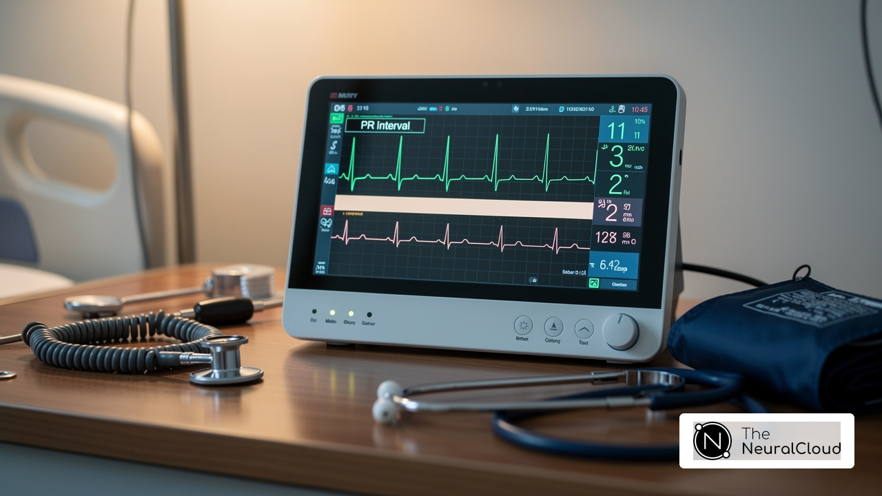
Electrocardiograms (ECGs) are essential graphical representations of the heart's activity, featuring the waveforms, including the P wave, QRS complex, and T wave. Each of these elements corresponds to specific electrical events within the heart:
- P Wave: This wave signifies atrial depolarization, indicating atrial contraction. Its normal amplitude is approximately 0.25 mV, with a duration ranging from 80 to 110 milliseconds.
- QRS Complex: This complex reflects ventricular depolarization, marking the rapid contraction of the ventricles. The typical duration of the QRS complex is between 80 and 100 milliseconds, with an R peak amplitude of about 1.6 mV.
- T Wave: The T wave represents ventricular repolarization, the recovery phase of the ventricles after contraction. Its duration typically lasts from 0.05 to 0.25 seconds, with an amplitude ranging from 0.1 to 0.8 mV.
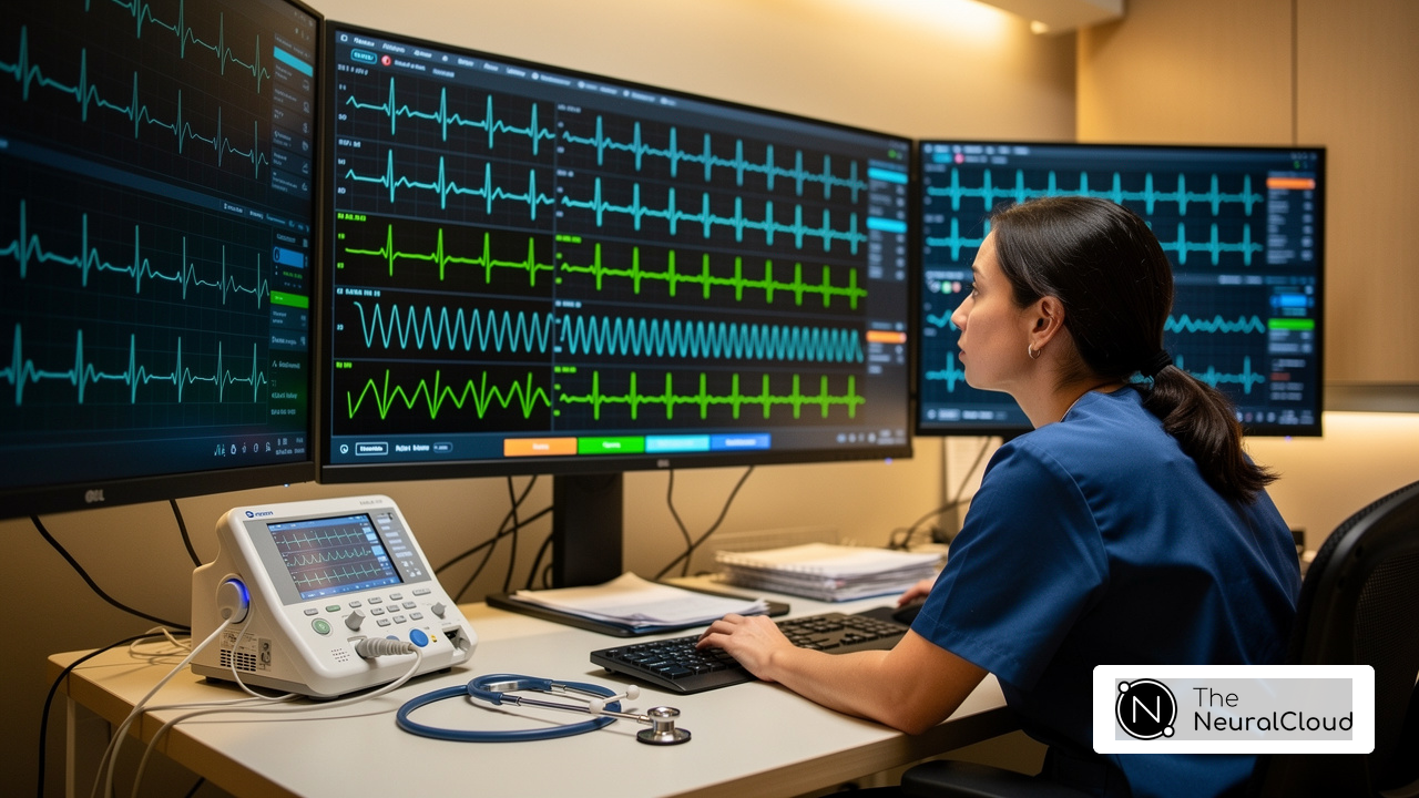
For health tech developers, understanding the basics of ECG waveforms is crucial for developing algorithms that effectively analyze ECG data. Recognizing the significance of each element allows programmers to create systems that operate with enhanced precision. The solutions from Neural Cloud Solutions offers insights and automation, improving workflow and addressing challenges such as physiological variability and noise. Expert insights highlight that interpretation of ECG is vital for diagnosing conditions, including arrhythmias and myocardial infarction. By mastering the intricacies of ECG waveforms and leveraging AI-driven automation, developers can significantly advance heart health monitoring and diagnostics.

Analyze Key Components: P-Wave, QRS Complex, and T-Wave
Each component of the waveforms of ECG is vital for cardiac function.
The P-Wave is typically small and rounded, indicating atrial depolarization. It is crucial for assessing atrial enlargement or other abnormalities. Research indicates that an enlarged P-Wave is associated with a two-fold risk of developing atrial fibrillation (AF), highlighting its importance in risk stratification.
The QRS Complex is characterized by its sharp, tall peaks, representing the rapid depolarization of the ventricles. Analysis of the QRS Complex can help identify conditions such as bundle branch blocks or ventricular hypertrophy. For instance, a prolonged QRS duration is necessary for diagnosing bundle branch block, while patients with QRS duration ≥150 ms often exhibit larger left ventricular volumes and lower ejection fractions.
The T-Wave is usually asymmetrical and reflects the repolarization of the ventricles. Changes in the T-Wave can indicate ischemia or electrolyte imbalances, which are critical for diagnosing various cardiac conditions.
By understanding the components of the ECG, creators can improve diagnostic accuracy, ultimately enhancing patient care.

Implement Advanced Techniques for Accurate ECG Interpretation
To achieve accurate ECG interpretation, developers can implement several techniques:
- Machine learning: Leveraging machine learning significantly enhances the classification and interpretation of ECG signals. By training algorithms on extensive datasets, developers enable systems to recognize patterns associated with various heart conditions, thereby improving diagnostic precision. For instance, studies have demonstrated high accuracy in detecting atrial fibrillation and other heart conditions, showcasing their practical applications in real-world scenarios.
- Advanced filtering methods: Advanced filtering methods are essential for reducing noise and artifacts in ECG signals, enhancing data clarity. Techniques such as wavelet transforms and adaptive filtering have proven particularly effective in isolating true cardiac signals from background noise, thus enhancing the accuracy for more reliable readings. Recent advancements, including a new algorithm that combines two-event related moving-averages (TERMA) and fractional-Fourier-transform (FrFT), further improve the analysis of ECG signals.
- Feature extraction: Identifying critical features from the ECG signals, such as intervals and amplitudes, enables more precise analysis. Automated processes streamline the process and minimize the risk of human error, leading to more consistent results. For example, high-resolution ECG techniques have effectively detected subtle changes indicative of myocardial ischemia, underscoring the importance of accuracy in clinical settings.
- AI-driven tools: Incorporating AI-driven tools facilitates real-time analysis and interpretation of ECG data, providing clinicians with immediate insights that can inform critical decision-making. This integration enhances the overall efficiency of ECG analysis, allowing for timely interventions in patient care. Statistics indicate that AI systems can achieve a negative predictive value of 99.2% for detecting acute conditions, emphasizing their effectiveness in clinical applications.
By leveraging these techniques, developers can create robust ECG analysis tools that significantly enhance diagnostic capabilities and improve patient outcomes. Additionally, user manuals and case studies demonstrating the application of these techniques using the MaxYield™ platform can provide further insights into their practical implementation.

Conclusion
Understanding the intricacies of ECG waveforms is essential for health tech developers seeking to enhance cardiac monitoring and diagnostics. By mastering the P wave, QRS complex, and T wave, developers can create more effective algorithms that accurately interpret these critical elements of heart activity. This knowledge not only facilitates improved risk assessment and diagnosis of heart conditions but also supports the development of innovative solutions in health technology.
Each waveform contributes significantly to a comprehensive understanding of cardiac health. The clinical significance of features, such as the P wave's role in atrial depolarization and the QRS complex's implications for ventricular function, cannot be overstated. Furthermore, integrating advanced techniques like machine learning and signal processing can significantly enhance the accuracy of ECG interpretation, enabling developers to create tools that assist healthcare professionals in making informed decisions.
Ultimately, mastering ECG waveforms and applying cutting-edge technologies are pivotal in advancing heart health monitoring. By embracing these insights and techniques, health tech developers can play a crucial role in transforming cardiac care, leading to improved patient outcomes and more effective interventions. The potential for innovation in this field is vast, and the time to act is now.
Frequently Asked Questions
What is an electrocardiogram (ECG)?
An electrocardiogram (ECG) is a graphical representation of the heart's electrical activity, depicting various waveforms that correspond to specific electrical events within the heart.
What are the main components of an ECG waveform?
The main components of an ECG waveform are the P wave, QRS complex, and T wave, each representing different phases of the heart's electrical activity.
What does the P wave signify in an ECG?
The P wave signifies atrial depolarization, indicating the contraction of the atria. Its normal amplitude is approximately 0.25 mV, with a duration ranging from 80 to 110 milliseconds.
What does the QRS complex represent in an ECG?
The QRS complex reflects ventricular depolarization, marking the rapid contraction of the ventricles. The typical duration of the QRS complex is between 80 and 100 milliseconds, with an R peak amplitude of about 1.6 mV.
What is the significance of the T wave in an ECG?
The T wave represents ventricular repolarization, which is the recovery phase of the ventricles after contraction. Its duration typically lasts from 0.05 to 0.25 seconds, with an amplitude ranging from 0.1 to 0.8 mV.
Why is understanding ECG waveforms important for health technology creators?
Understanding ECG waveforms is crucial for health technology creators as it enables them to develop algorithms that effectively analyze ECG data, leading to enhanced precision in heart signal analysis.
What features does the MaxYield™ platform offer for ECG analysis?
The MaxYield™ platform from Neural Cloud Solutions offers advanced noise filtering and automation, improving workflow and addressing challenges such as physiological variability and signal artifacts in ECG data.
How does accurate identification of ECG waveforms impact heart health diagnostics?
Accurate identification of ECG waveforms is vital for diagnosing various heart conditions, including arrhythmias and myocardial infarction, allowing for better heart health monitoring and diagnostics.
List of Sources
- Explore the Basics of ECG Waveforms
- researchgate.net (https://researchgate.net/publication/327110331_ECG_Waveform_Classification_Based_on_P-QRS-T_Wave_Recognition)
- Normal Electrocardiography (ECG) Intervals: Normal Electrocardiography Intervals (https://emedicine.medscape.com/article/2172196-overview)
- nature.com (https://nature.com/articles/s41586-025-09227-0)
- escardio.org (https://escardio.org/Councils/Council-for-Cardiology-Practice-(CCP)/Cardiopractice/artificial-intelligence-in-ecg-diagnostics-where-are-we-now)
- pmc.ncbi.nlm.nih.gov (https://pmc.ncbi.nlm.nih.gov/articles/PMC7664289)
- Analyze Key Components: P-Wave, QRS Complex, and T-Wave
- pmc.ncbi.nlm.nih.gov (https://pmc.ncbi.nlm.nih.gov/articles/PMC9714955)
- QRS Interval (https://litfl.com/qrs-interval-ecg-library)
- ahajournals.org (https://ahajournals.org/doi/10.1161/CIRCEP.121.010435)
- pmc.ncbi.nlm.nih.gov (https://pmc.ncbi.nlm.nih.gov/articles/PMC5292639)
- Checking your browser - reCAPTCHA (https://pmc.ncbi.nlm.nih.gov/articles/PMC6231906)
- Implement Advanced Techniques for Accurate ECG Interpretation
- nature.com (https://nature.com/articles/s41598-021-97118-5)
- escardio.org (https://escardio.org/Councils/Council-for-Cardiology-Practice-(CCP)/Cardiopractice/artificial-intelligence-in-ecg-diagnostics-where-are-we-now)
- Current and Future Use of Artificial Intelligence in Electrocardiography - PMC (https://pmc.ncbi.nlm.nih.gov/articles/PMC10145690)
- medicalsearch.com.au (https://medicalsearch.com.au/buying-guide/advancements-in-ecg-technology-and-signal-processing/f/24927)
- Client Challenge (https://nature.com/articles/s41591-023-02396-3)