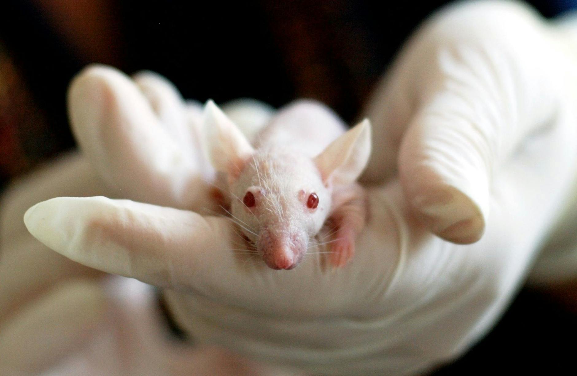
Mouse models have become the foundation of preclinical cardiac research. Scientists prefer mice because their heart biology is easy to study, they reproduce rapidly, and their genetics are well understood. This makes them perfect for testing new ideas before moving to larger animals or people.
The main goal of using mice data is to better predict how new treatments will work in human hearts. By carefully modelling heart attacks, heart failure, and other cardiac conditions in mice, researchers can explore everything from how heart attacks heal to the ways immune cells help or harm recovery. Reliable mouse data leads to stronger evidence and smoother paths from the lab to new therapies.
Why Mice are Critical in Cardiac Preclinical Research
Mice are not just small, furry creatures; in the lab, they are powerful tools for understanding the heart. Their size, short life cycle, and advanced genetic tools make them a favorite among cardiac researchers. When we want to predict how a drug or therapy will affect human hearts, mouse data often forms our starting point. Mice help us connect new science to practical treatments, making progress faster and more reliable.
Genetic Manipulability and Transgenic Models
One major reason mice shine in preclinical cardiac research is their genetic flexibility. Scientists can change, knock out, or replace specific genes in mice with tools that are much harder to use in other animals. This lets us mimic human heart diseases right at the genetic level.
Common genetically modified mouse models include:
- LDLR−/− mice: Lacking the LDL receptor gene, these mice develop high cholesterol and plaques in their arteries. They help researchers study atherosclerosis, which is a leading cause of heart attacks in people.
- apoE−/− mice: These mice miss the gene for apolipoprotein E, causing them to develop cholesterol-rich plaques rapidly, making them valuable for testing new drugs to lower cholesterol or stop artery disease.
- apoE3Leiden mice: Carrying a human-like mutation, these mice have a fat metabolism close to some people, providing an even better match for studying certain types of heart disease.
These models allow scientists to study how a single genetic change can ripple out and affect the whole cardiovascular system. This has paved the way for discovering what drives heart failure, high cholesterol, and even rare heart muscle diseases. For a deeper look on how these genetic tools impact cardiac studies, read this overview on using mouse genetics to study cardiac hypertrophy and heart disease.
Physiological and Anatomical Relevance to Human Cardiac Disease
How much do mice really reflect the human heart? While there are important similarities, there are also some big differences. Their hearts are made up of the same basic structures — valves, chambers, arteries, and the same beating and pumping action we depend on.
Both mouse and human hearts:
- Respond to stress by growing (hypertrophy)
- Develop similar plaque buildups in arteries under the right genetic conditions
- Show parallel patterns during heart injury and repair
But, we must keep in mind a few important differences:
- Size and heart rate: A mouse’s heart beats 500 to 600 times per minute, much faster than a human’s 60 to 100. This changes the way their heart muscle responds during stress or disease.
- Lifespan and metabolism: Mice live for 1 to 2 years and process drugs and nutrients more quickly than people.
- Genetic variation: Mice bred in labs can be very homogeneous, making them good for comparing results but sometimes less representative of the wide genetic variation in humans.
So, while they share a strong foundation, careful interpretation is critical. Want more details on how researchers measure and interpret mouse heart function? Visit the Guidelines for measuring cardiac physiology in mice.
Advantages and Limitations of Mouse Models
With all these benefits, it’s easy to forget that mice aren’t perfect stand-ins for people. Researchers value them for a mix of reasons:
Main advantages:
- Cost-effective: Mice are less expensive to buy, house, and care for than larger animals.
- Ease of handling: Their small size and adaptability make mice easy to work with in a lab.
- Genetic tools: Decades of genetic work mean scientists can easily buy or create mice with the exact changes they need.
- High-throughput studies: Many mice can be studied at once, helping speed up research.
Main limitations:
- Species differences: As mentioned, their rapid heart rate and distinct metabolism can make some results hard to translate directly to people.
- Scale: What works in a tiny mouse heart may not always scale up to work in a human heart.
- Complex human diseases: Mice sometimes fail to capture the full range of triggers and symptoms seen in human patients.
Choosing the right mouse model means balancing the benefits against these challenges. Researchers weigh these points every time they set up an experiment or interpret data. For more perspective, check this review on animal models of cardiovascular diseases and the discussion of advantages and limitations in cardiac research.
Mice remain the first choice because their strengths match what early-stage research needs: reliable genetics, manageable costs, and a bridge between isolated lab work and meaningful studies in people.
Methodologies for Cardiac Phenotyping and Data Collection in Mice
Mouse data drives progress in cardiac research, but the way we measure and analyze heart function in mice matters as much as the models themselves. Reliability starts with the tools and approaches researchers use to spot subtle changes in heart size, motion, and pressure, then track disease as it develops or heals. Here’s how experts gather and interpret this complex data to fuel new discoveries.
Invasive and Non-Invasive Cardiac Assessment Techniques
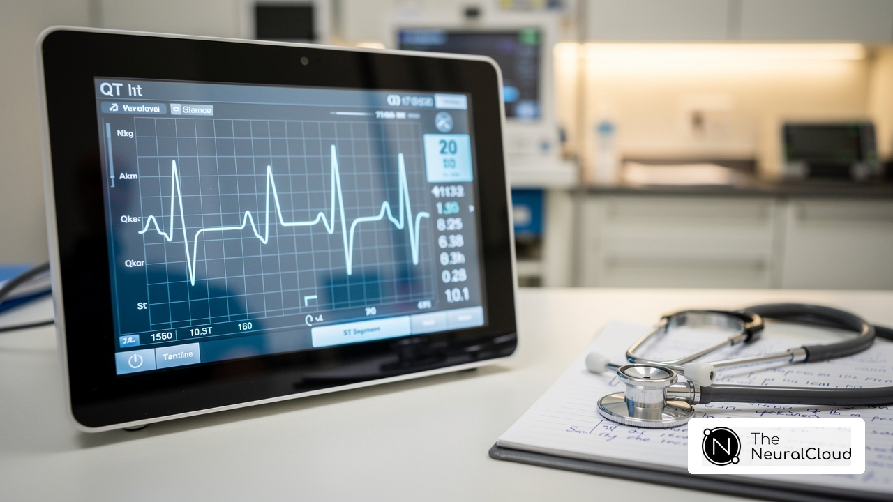
Mice may be small, but the tools for studying their hearts rival those used in clinical medicine. Scientists use both invasive and non-invasive methods, each with their own benefits and hurdles.
- Echocardiography (Echo):
- This high-frequency ultrasound imaging lets researchers see the mouse heart in real-time. It’s painless, non-invasive, and used for tracking heart chamber size, wall thickness, and how well the heart pumps.
- 3D and Doppler echocardiography add even more detail, measuring flow and subtle deformities.
- Technical challenges include the need for skilled operators, high-resolution equipment, and managing the fast mouse heart rate. Learn more about non-invasive cardiac output measurement in mice.
- Speckle-Tracking Imaging:
- A newer advance on standard echo, this technique tracks small patterns (“speckles”) in the heart muscle as it moves. This reveals early signs of dysfunction before changes show up in conventional scans.
- Fast imaging speeds and advanced software are essential, since the mouse heart beats so quickly.
- Cardiac MRI (Magnetic Resonance Imaging):
- Cardiac MRI gives razor-sharp images of the mouse heart in any plane. It’s the gold standard for measuring volumes, scar tissue, and perfusion.
- MRI is powerful, but equipment is costly and the scans take longer, requiring mice to stay perfectly still (often under anesthesia).
- Invasive Pressure-Volume (PV) Loop Analysis:
- With a tiny catheter inside the heart, this method measures real-time changes in pressure and volume through the heartbeat. It’s the best way to understand cardiac contractility and stiffness.
- Invasive PV loops are sensitive but technically demanding, requiring surgical skill and specialized catheters. Newer non-invasive approaches are being validated, offering promise for future studies (see this recent paper on non-invasive PV loop methods).
- Choosing the right tool: The choice depends on what’s being measured—structure, function, or hemodynamics—as well as lab resources and expertise. Echo offers speed and convenience, MRI offers unmatched detail, while PV analysis digs deepest into mechanical function. For a broader look at the toolbox, check out this review on non-invasive assessment of heart failure in mouse models.
Disease Models and Experimental Protocols
Rigorous, reproducible data starts with the right model and protocol. Cardiac disease doesn’t look the same in every mouse—or every experiment—unless protocols are clear and consistent.
- Myocardial Infarction (Heart Attack) Models:
- Coronary artery ligation is the go-to method. A tiny suture ties off a main coronary artery, causing a region of the heart to lose blood supply and mimicking a heart attack.
- Ischemia-Reperfusion Injury:
- Here, the artery is blocked for a short window, then blood flow is restored. This models the injury that occurs both during and after a heart attack—critical for testing new therapies.
- Pressure Overload (TAC, or Transverse Aortic Constriction):
- TAC uses a surgical band to narrow the aorta, pushing the heart to work harder. Over time, mice develop heart muscle thickening and heart failure that closely parallels human disease.
- Atherogenesis and Heart Failure Models:
- High-fat diets and genetic backgrounds (like LDLR−/− and apoE−/−) encourage plaque formation, while different stressors—like TAC or doxorubicin—trigger heart failure pathways.
- Protocol Standardization and Collaboration:
- Consistency matters. Labs now use shared guidelines, detailed methods, and multicenter trial designs to cut down on errors and bias. This boosts the odds that results are true, not just interesting.
- Researchers often work together across sites, using shared data repositories and harmonized protocols. This has led to more repeatable, reliable findings—key for translation to human therapies. For specific methods and examples, dive into experimental rodent models of cardiovascular diseases, and expand your knowledge with this review of animal models in cardiovascular research.
Emerging Technologies and Data Analysis Approaches
New tools don’t just make measurements sharper—they make mouse cardiac studies faster, more accurate, and easier to compare across labs.
- Imaging Advances:
- Virtual histology and advanced MRI sequences let scientists see heart tissue changes in fine detail.
- Hybrid methods (using PET/CT or PET/MR) give both anatomical and molecular insights, connecting structure with processes like inflammation or scarring. For current directions, see biomedical imaging advances in experimental models.
- Digital Data Analysis Tools:
- AI and machine learning now help process imaging data, spot patterns, and predict outcomes from vast datasets. These tools make it possible to study small changes, find trends, and generate reproducible results across cohorts. Curious how artificial intelligence is changing cardiovascular analysis? Visit this recent review on AI in cardiovascular medicine.
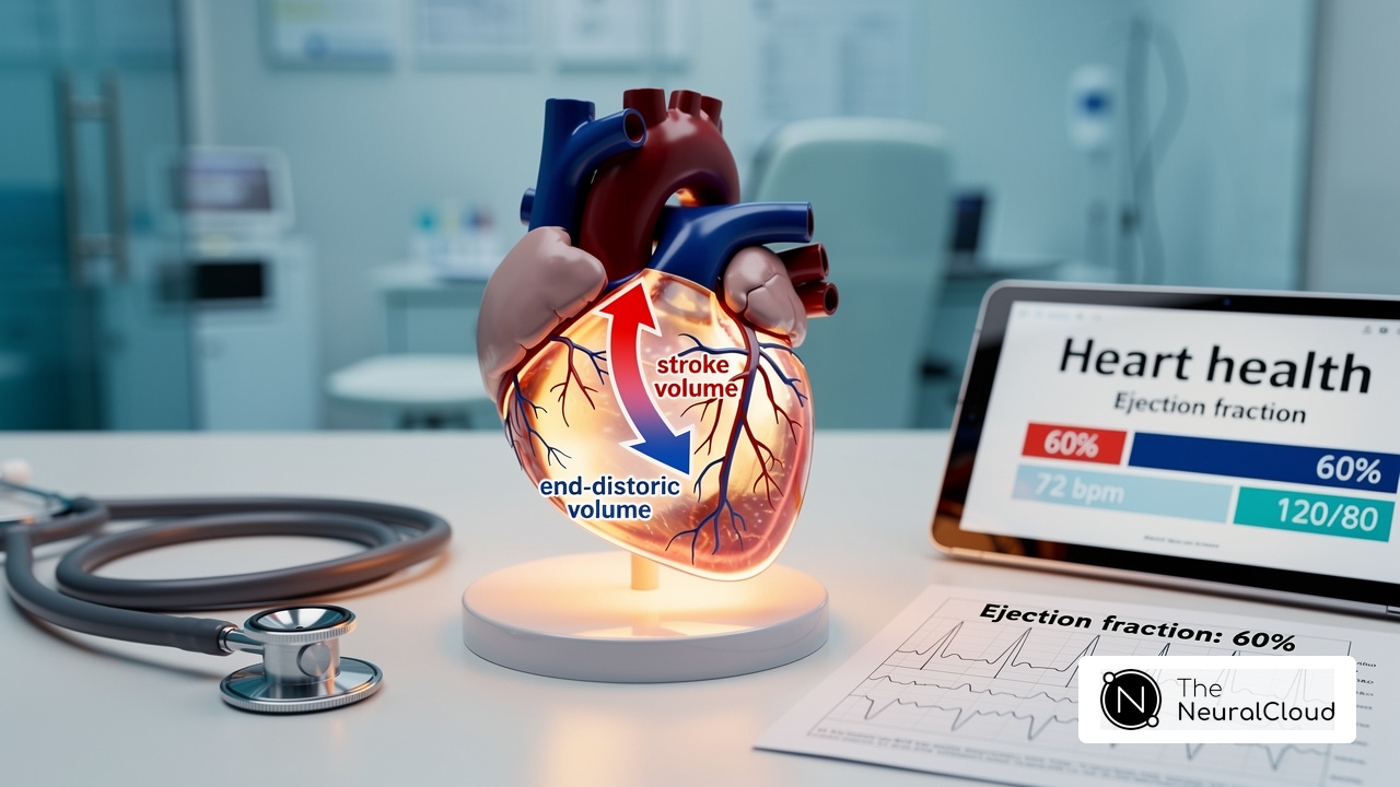
- Software can now automate ejection fraction calculations, wall thickness measurements, and even predict which mice may develop severe disease.
- Standardizing Data for Translational Research:
- Open-source data platforms and digital lab notebooks mean researchers can store, share, and review data with more oversight and transparency.
- Multicenter studies using these platforms increase trust in published work, highlight outliers, and promote the best practices globally. They pave the way for new discoveries to move smoothly from animal studies to human trials, as discussed in strategies for cardiac preclinical research and data reproducibility.
- Collaborative Networks:
- Big research networks help set data standards, pool results, and speed up the hunt for new heart therapies. This teamwork is the new normal for cardiac research and pushes the whole field forward.
As new technology rolls out and collaboration broadens, mouse data becomes even more valuable. This progress is making preclinical cardiac studies more predictive and linking results more tightly to what patients actually experience.
Translational Value and Future Directions of Mice Data in Cardiac Research
Mice data are the linchpin for progress in preclinical cardiac research, but the true test is whether results hold up when new therapies move from bench to bedside. Knowing what works in a mouse heart is only the first step; what matters most is how reliably those discoveries turn into benefits for patients. This section covers where mouse-based breakthroughs have paved the way for treatments, the challenges that hold back translation, and the advances poised to boost the value of mouse data for human heart care.
Case Studies of Therapeutic Discovery and Validation
Several treatments now offered to cardiac patients owe much of their story to mouse model discoveries. These case studies highlight the path from animal findings to real-world impact.
- PCSK9 Inhibitors: Researchers first uncovered the PCSK9 gene’s role in cholesterol control through mouse studies, leading to the development of drugs now widely used to help people lower stubborn LDL cholesterol and reduce heart attack risk.
- Statins: Genetic mouse models that develop arterial plaques (like apoE−/− and LDLR−/− mice) were crucial in understanding how statins slow atherosclerosis.
- SGLT2 Inhibitors: Originally diabetes drugs, SGLT2 inhibitors were observed to improve heart function in mouse models of heart failure, setting the stage for successful large-scale human trials. These drugs now help both diabetic and non-diabetic heart failure patients.
- Stem Cell and Gene Therapies: Mouse experiments helped uncover how cell therapies or gene editing might repair damaged hearts after a heart attack, with several approaches now in early-phase human trials.
Consider the formation of databases like the Mouse Heart Attack Research Tool, which pool massive datasets and help validate cardiac findings across centers before moving to clinical development. This data-driven approach has helped filter out less promising treatments early, freeing up resources for those with clear translational promise.
Challenges in Translating Mouse Data to Human Cardiology
Success stories aside, translating results from mice to people doesn’t always work as planned. Many therapies that look promising in mouse models fail to deliver the same benefits for human hearts.
Some of the toughest challenges include:
- Species Differences: Mice and humans differ in heart rate, physiology, immune system response, and the way their bodies handle drugs. For example, mice heal heart injury differently, often showing less scarring and quicker cell turnover compared to people.
- Genetic Uniformity: Laboratory mice are often genetically similar, much more so than human populations. This makes it hard to predict how therapies will work in a diverse group of patients.
- Disease Complexity: Human cardiac disease often involves multiple factors (age, environment, genetics, diet), while mouse models may isolate only one feature at a time.
- Regulatory Hurdles: Agencies now expect robust, reproducible animal data from multiple labs before giving approval to start human trials. A lack of standardization in mouse experiments can slow the entire process.
- Poor Predictive Value: Studies have shown weak correlation between some genomic responses in mouse models and humans after cardiac injury, leading to a re-examination of what preclinical success means (more on these gaps).
- Bias and Reporting Issues: Inadequate blinding and selective reporting in some mouse studies can overstate findings and inflate the chances of a failed human trial.
A deeper dive into these topics is available in this overview of limitations in mouse models for cardiovascular research, which highlights both the useful similarities and the mismatches that still need solutions.
Opportunities for Improving Reproducibility and Translational Success
The scientific community recognizes that better reproducibility and careful study design are the keys to unlocking more successful human therapies based on mouse research. Ongoing efforts are raising the bar for how data is collected and shared.
- Standardized Protocols: Using shared guidelines and step-by-step protocols across labs reduces variability, making results more reliable. The adoption of standards for everything from anesthesia to imaging helps ensure findings can be trusted and replicated elsewhere.
- Multicenter Preclinical Trials: By pooling resources and running studies across multiple labs, researchers can spot inconsistencies early, improve data quality, and build a foundation that mimics large human studies.
- Data Sharing Platforms: Open-access databases and collaborative tools make it easier to share raw and processed data. This transparency allows for broader validation and can quickly identify errors or unexpected patterns, leading to fewer surprises in clinical trials.
- Refined Statistical Methods: Improved statistical planning, inclusion of both sexes, and rigorous blinding and randomization cut down on the risk of bias and enhance the reliability of findings. Major journals increasingly require clear reporting on study design, animal numbers, and reproducibility measures.
- Improving Rigor and Reproducibility Initiatives: Groups are developing specific best practices, such as those outlined in improving rigor and reproducibility in cardiovascular research and practical guides like these recommendations for cardiovascular studies.
- Encouraging Methodological Rigor: Thoughtful discussion about the need for careful experimental design, as highlighted in the article methodological rigor in preclinical cardiovascular studies, pushes the field to commit to higher standards.
These efforts are not only making mouse research more credible, they’re building bridges so that the best preclinical discoveries have a real shot at becoming tomorrow’s cardiac therapies. As standards rise and cooperation grows, the gap between animal studies and patient treatment continues to narrow, giving hope for smoother and more reliable progress in cardiac care.
Conclusion
Mice data continue to anchor progress in preclinical cardiac research, helping scientists test new ideas quickly and clearly. When researchers use careful protocols and share data across labs, their findings become more reliable for building tomorrow’s therapies. This ongoing teamwork, backed by new technology and smarter standards, means studies can predict human success with greater confidence.
With every advance, the link between mouse models and human treatments grows stronger. Staying focused on rigorous methods and open collaboration will keep moving the field forward, opening doors for faster breakthroughs and better heart care. Thanks for reading—share your thoughts or experiences with mouse models and let’s keep the conversation going about making cardiac research more reliable and innovative.