Overview
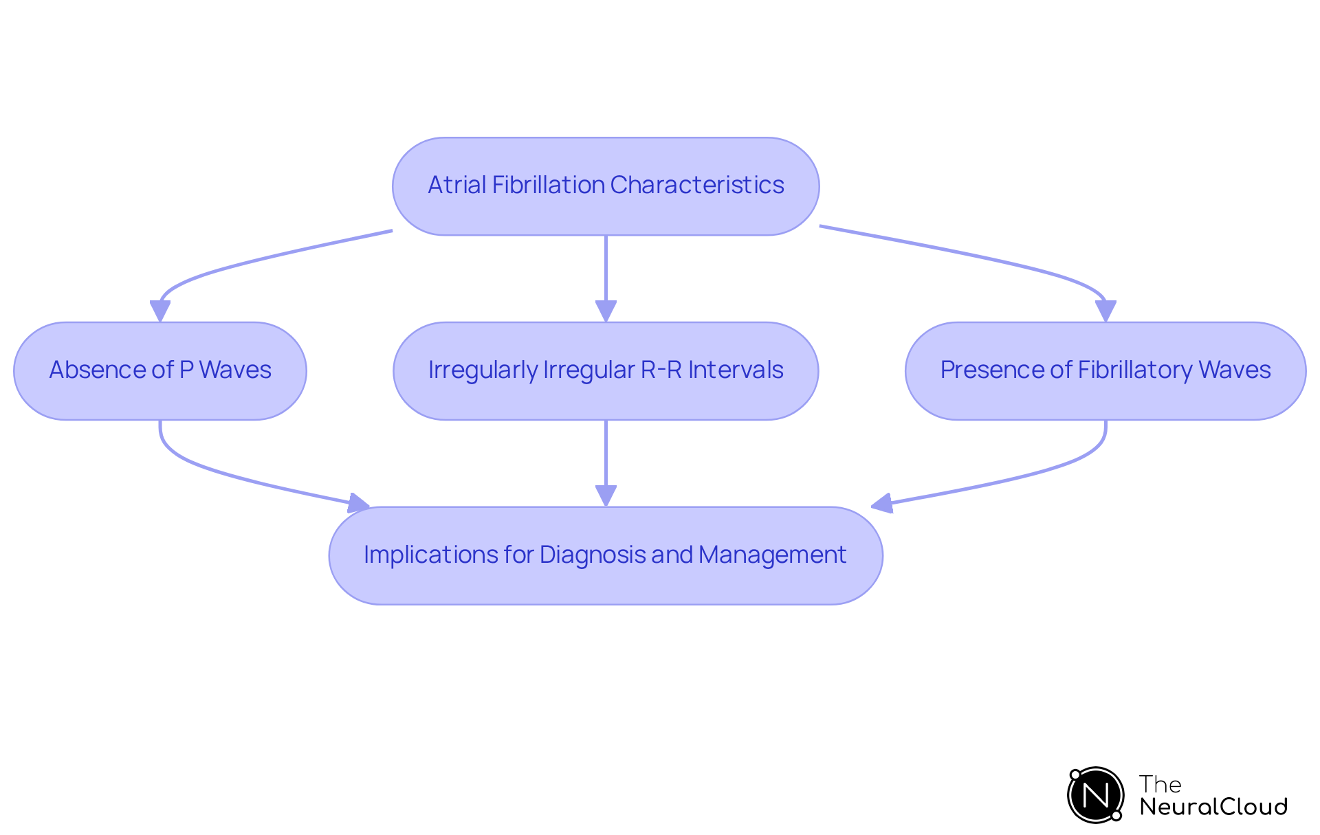
Atrial fibrillation (Afib) on an EKG presents distinct characteristics that are crucial for diagnosis. Key features include:
- The absence of distinct P waves
- Irregularly irregular R-R intervals
- The presence of fibrillatory waves
These indicators reflect chaotic electrical activity in the atria, which can lead to significant health risks such as stroke and heart failure.
Recognizing these features is essential for effective management of Afib. Accurate EKG interpretation plays a vital role in identifying patients at risk, allowing healthcare professionals to implement timely interventions. Moreover, the integration of advanced technologies can enhance detection capabilities, ensuring that patients receive appropriate care.
In summary, understanding the nuances of Afib on an EKG not only aids in diagnosis but also contributes to better patient outcomes. By prioritizing accurate interpretation and leveraging technological advancements, healthcare providers can significantly reduce the risks associated with this condition.
Introduction
Understanding how atrial fibrillation (AFib) appears on an electrocardiogram (EKG) is crucial, particularly as this arrhythmia's prevalence continues to rise, impacting millions. This article explores the unique characteristics of AFib on EKGs, notably the absence of P waves and the irregularity of R-R intervals that indicate this condition.
Given the complexities of EKG readings and the risk of misdiagnosis, how can healthcare professionals ensure accurate identification and timely intervention? By examining the nuances of AFib on EKGs, we can enhance diagnostic accuracy, which significantly influences patient outcomes, making this knowledge vital in clinical practice.
Define Atrial Fibrillation and Its EKG Representation
Atrial fibrillation is a common arrhythmia characterized by rapid and irregular beating of the heart's upper chambers, known as the atria. To understand what does afib look like on an ekg, it is identified by the absence of distinct P waves, which are typically present in normal sinus rhythm. Instead, the EKG shows irregularly irregular R-R intervals and chaotic electrical activity in the atria, often referred to as fibrillatory waves. This disorganized electrical activity leads to ineffective atrial contractions, significantly increasing the risk of thromboembolic events, including stroke.
Recent studies indicate a dramatic rise in the incidence of atrial fibrillation, with estimates suggesting that approximately 10 million Americans are affected. Notably, after the age of 40, 1 in 4 individuals will develop atrial fibrillation, and 1 in 5 will face heart failure. The CHA2DS2-VASc score is frequently utilized to evaluate stroke risk in individuals with atrial fibrillation, guiding treatment decisions.
Key characteristics of atrial fibrillation on EKG include:
- Absence of P waves
- Irregularly irregular R-R intervals
- Presence of fibrillatory waves
Research has shown that patients with atrial fibrillation who undergo surgical ablation during cardiac procedures experience improved long-term survival and quality of life. This underscores the importance of identifying what does afib look like on an ekg and addressing it promptly to mitigate associated risks. Experts emphasize that understanding what does afib look like on an ekg is crucial for effective diagnosis and management, especially in patients undergoing cardiac surgery, where treatment is often overlooked despite established guidelines.
Case studies highlight the significance of understanding what does afib look like on an ekg in diagnosing atrial fibrillation. For instance, a systematic review found that P-wave duration is a distinct predictor of atrial fibrillation recurrence following catheter ablation, emphasizing the need for accurate EKG interpretation in clinical practice. Moreover, advancements in wearable technology, such as smartwatches, are facilitating the early detection of atrial fibrillation, enabling timely intervention and improved patient outcomes.
Explain the Clinical Importance of Identifying Afib on EKGs
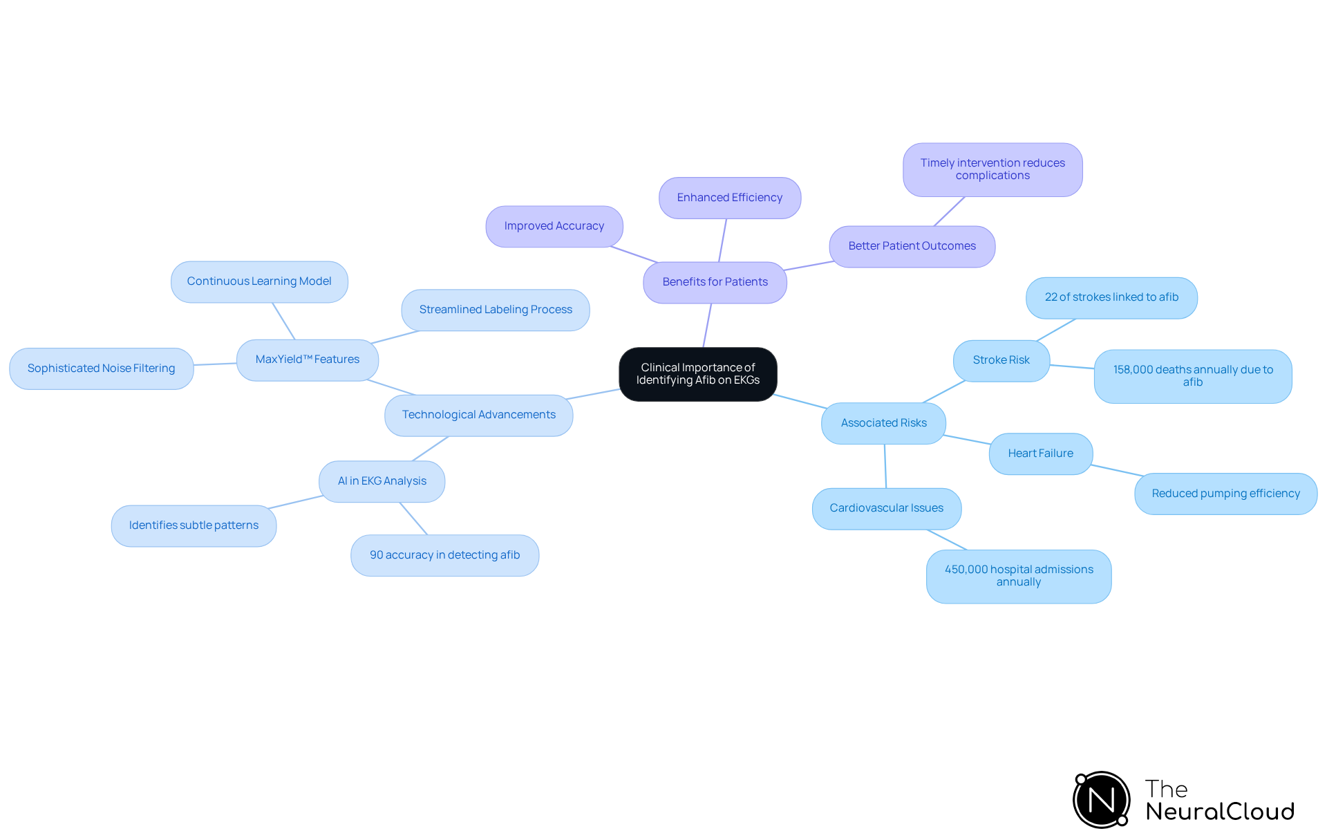
Recognizing what does afib look like on an ekg is essential due to its association with increased risks of stroke, heart failure, and other cardiovascular issues. Approximately 22 percent of strokes are linked to atrial fibrillation, underscoring the importance of early detection for prompt intervention. This may include anticoagulation therapy, which can significantly lower stroke risk. Furthermore, identifying atrial fibrillation enables clinicians to customize treatment plans, including rate or rhythm control strategies, ultimately improving patient outcomes.
Recent advancements in digital health technologies, particularly AI-enabled EKG analysis, have improved the accuracy of atrial fibrillation detection. Research indicates that AI can identify subtle patterns of atrial fibrillation with up to 90 percent accuracy, even amidst regular heart rhythms. This capability is crucial, given that atrial fibrillation results in over 450,000 hospital admissions annually in the U.S. and contributes to around 158,000 deaths each year.
Current guidelines from leading cardiology associations emphasize the need for early identification and treatment of atrial fibrillation, which includes knowing what does afib look like on an ekg. For instance, the 2023 ACC/AHA/ACCP/HRS guidelines advocate for proactive strategies in diagnosing and managing atrial fibrillation, emphasizing the importance of knowing what does afib look like on an ekg to mitigate its progression and associated risks. Real-world examples, such as the use of digital health tools, illustrate improved outcomes for patients through early detection and intervention strategies.
Expert insights highlight that early recognition of atrial fibrillation symptoms is vital for effective management. By utilizing advanced technologies like Neural Cloud Solutions' MaxYield™, which features sophisticated noise filtering and a continuous learning model, healthcare professionals can enhance the accuracy and efficiency of ECG analysis. MaxYield™ streamlines the labeling process, addressing challenges such as physiological variability and signal artifacts, ultimately improving the quality of care for patients with atrial fibrillation and reducing the risk of severe complications like stroke.
Features of MaxYield™
- Sophisticated Noise Filtering: Enhances signal clarity for more accurate readings.
- Continuous Learning Model: Adapts to new data, improving analysis over time.
- Streamlined Labeling Process: Reduces time spent on ECG interpretation.
Advantages for Healthcare Professionals
- Improved Accuracy: Higher detection rates of atrial fibrillation.
- Enhanced Efficiency: Saves time in ECG analysis, allowing for quicker patient management.
- Better Patient Outcomes: Reduces the likelihood of complications through timely intervention.
Describe Key EKG Features of Atrial Fibrillation
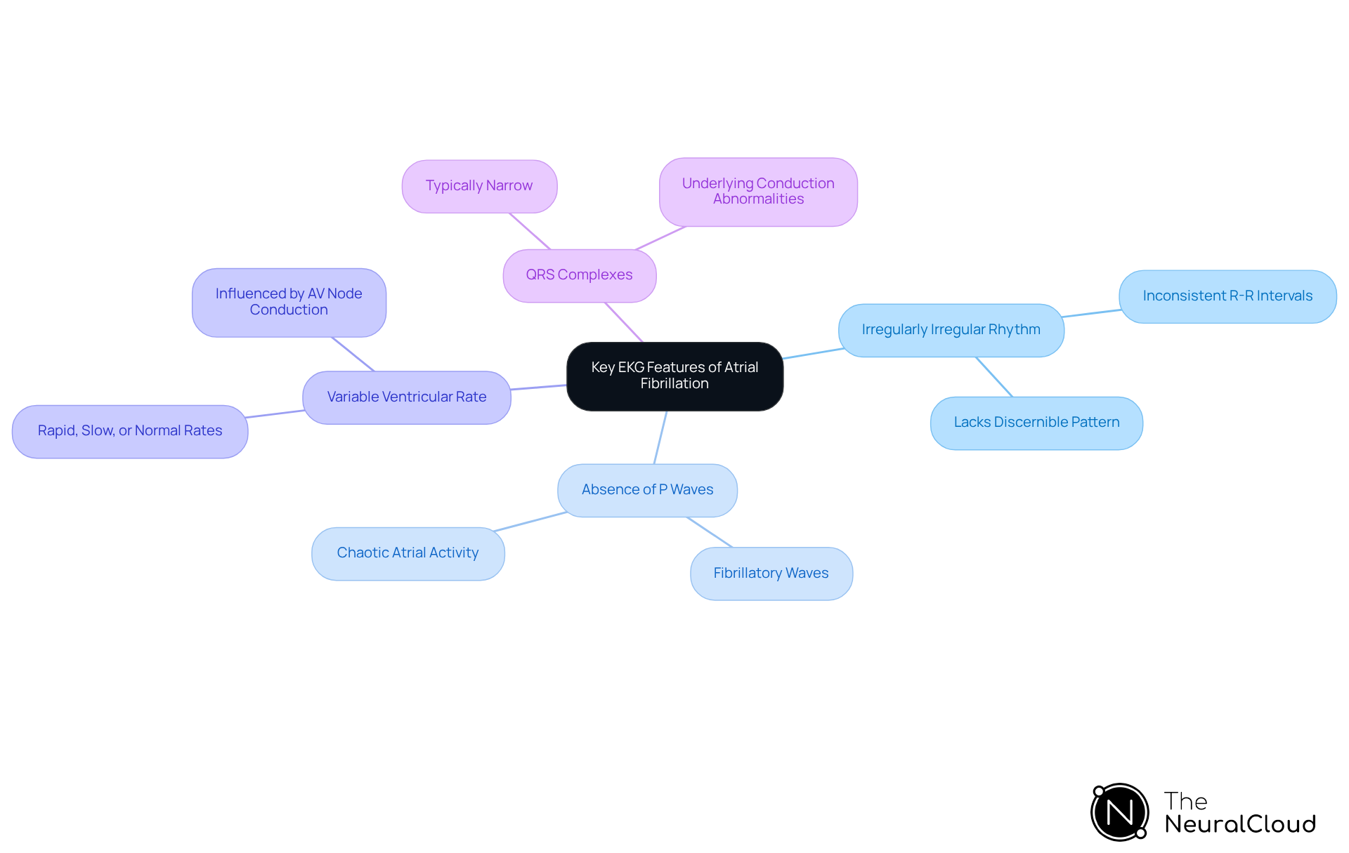
Understanding what does afib look like on an ekg is critical for precise diagnosis and management. Key characteristics include:
- Irregularly Irregular Rhythm: The R-R intervals exhibit inconsistency, lacking a discernible pattern, which is a defining trait of AFib.
- Absence of P Waves: Instead of the typical P waves, fibrillatory waves may be observed, varying in amplitude and frequency, indicating chaotic atrial activity. This absence is a definitive sign of atrial fibrillation, which impacts around 6 million individuals in the U.S. and is linked to a considerable risk of stroke, as approximately 15 to 20 percent of stroke patients experience this condition.
- Variable Ventricular Rate: The ventricular response can fluctuate, presenting as rapid, slow, or normal rates, influenced by conduction through the AV node.
- QRS Complexes: Typically, the QRS complexes are narrow unless there is an underlying conduction abnormality.
These features collectively reflect the disorganized electrical impulses typical of atrial fibrillation. Significantly, atrial fibrillation is more prevalent after age 60, with 8% of individuals over 80 impacted. Understanding what afib looks like on an ekg is essential for healthcare professionals in diagnosing and managing this prevalent arrhythmia effectively.
To enhance the diagnostic process, healthcare professionals can leverage MaxYield™, an advanced AI-driven ECG analysis platform from Neural Cloud Solutions. MaxYield™ automates the mapping of ECG signals through noise, isolating and labeling key features in every heartbeat. This capability allows for rapid analysis of 200,000 heartbeats in less than 5 minutes, providing detailed insights that support confident clinical decision-making.
Features of MaxYield™:
- Automates ECG signal mapping
- Isolates and labels key features in each heartbeat
- Analyzes 200,000 heartbeats in under 5 minutes
Advantages for Healthcare Professionals:
- Enhances identification of atrial fibrillation characteristics
- Improves patient outcomes through rapid analysis
By incorporating MaxYield™ into their workflow, healthcare professionals can enhance the identification of atrial fibrillation characteristics, ultimately improving patient outcomes. As Dr. Nicholas Olson, a cardiac electrophysiologist, states, "Atrial fibrillation is a serious heart condition that increases the risk of stroke.
Clarify Misconceptions and Variations in Afib EKG Readings
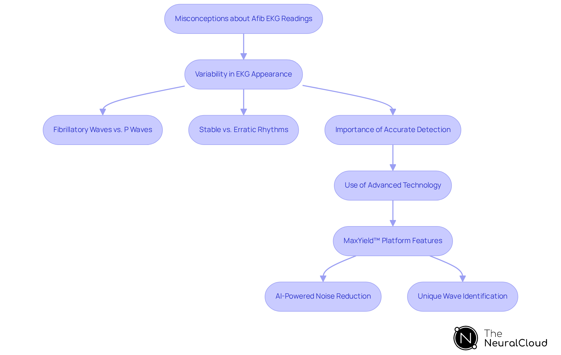
Misunderstandings regarding atrial fibrillation often stem from the variability in what does afib look like on an ekg. The subtle distinction between fibrillatory waves and P waves can lead to potential misdiagnosis. Atrial fibrillation can manifest differently among individuals; some may exhibit a more erratic baseline, while others may have a comparatively stable rhythm. Additionally, atrial fibrillation can be paroxysmal, meaning it may occur intermittently, which complicates diagnosis if the EKG is not performed during an active episode. Statistics indicate that about 25% of individuals experiencing their first stroke show signs of atrial fibrillation, highlighting the critical need for accurate detection.
Understanding these nuances is essential for healthcare providers to ensure precise interpretation and effective patient management. Heart specialists emphasize that what does afib look like on an ekg is characterized by the presence of 'irregularly irregular' rhythms without distinct P waves, which is a hallmark of atrial fibrillation. This underscores the importance of comprehensive training in EKG analysis, especially in recognizing what does afib look like on an ekg, to prevent misdiagnosis and ensure timely treatment. The integration of advanced technologies, such as Neural Cloud Solutions' MaxYield™ platform, can significantly enhance this diagnostic process.
MaxYield™ offers several key features that improve ECG analysis:
- AI-Powered Noise Reduction: This feature minimizes background noise, allowing for clearer readings.
- Unique Wave Identification: It enhances the ability to distinguish between different waveforms, improving accuracy in detecting atrial fibrillation.
The advantages of using MaxYield™ for healthcare professionals are substantial. By enhancing the clarity of ECG recordings, the platform enables more precise detection of atrial fibrillation. This is crucial, as untreated AFib can lead to serious health consequences, including an increased risk of stroke and heart failure. The urgency of accurate diagnosis cannot be overstated, making tools like MaxYield™ invaluable in the healthcare landscape.
Conclusion
Atrial fibrillation is a significant cardiac condition that requires careful attention due to its potential complications, particularly the risk of stroke and heart failure. Recognizing the unique features of atrial fibrillation on an EKG—such as the absence of P waves and irregularly irregular R-R intervals—is crucial for timely diagnosis and effective management. Understanding the EKG representation of atrial fibrillation is essential for healthcare professionals, as it directly impacts patient outcomes and treatment strategies.
The article highlights several key points regarding the clinical importance of identifying atrial fibrillation on EKGs. Early detection can lead to timely interventions, such as anticoagulation therapy, which significantly reduces stroke risk. The integration of advanced technologies, including AI-driven tools like MaxYield™, enhances the accuracy of EKG analysis. This allows healthcare providers to detect subtle patterns of atrial fibrillation with greater confidence. Furthermore, addressing common misconceptions about EKG readings can prevent misdiagnosis and ensure that patients receive appropriate care.
In light of the rising prevalence of atrial fibrillation, it is imperative for healthcare professionals to remain vigilant in recognizing its EKG characteristics. By leveraging innovative diagnostic tools and continuing education on EKG interpretation, clinicians can improve patient care and outcomes. A proactive approach to identifying atrial fibrillation not only mitigates associated risks but also empowers patients to take charge of their heart health.
Key Features of MaxYield™:
- Enhanced Accuracy: AI-driven analysis improves detection of atrial fibrillation patterns.
- Timely Interventions: Facilitates early diagnosis, leading to prompt treatment decisions.
- User-Friendly Interface: Designed for ease of use by healthcare professionals.
Advantages for Healthcare Professionals:
- Improved Patient Outcomes: Early detection and treatment reduce the risk of complications.
- Informed Decision-Making: Access to accurate data supports better clinical decisions.
- Empowerment through Education: Continuous learning opportunities enhance EKG interpretation skills.
Frequently Asked Questions
What is atrial fibrillation?
Atrial fibrillation is a common arrhythmia characterized by rapid and irregular beating of the heart's upper chambers, known as the atria.
How can atrial fibrillation be identified on an EKG?
Atrial fibrillation can be identified on an EKG by the absence of distinct P waves, irregularly irregular R-R intervals, and the presence of chaotic electrical activity referred to as fibrillatory waves.
What are the risks associated with atrial fibrillation?
Atrial fibrillation leads to ineffective atrial contractions, significantly increasing the risk of thromboembolic events, including stroke.
How prevalent is atrial fibrillation among Americans?
Approximately 10 million Americans are affected by atrial fibrillation, with estimates suggesting that after the age of 40, 1 in 4 individuals will develop it.
What is the CHA2DS2-VASc score?
The CHA2DS2-VASc score is a tool used to evaluate stroke risk in individuals with atrial fibrillation, guiding treatment decisions.
What are the key characteristics of atrial fibrillation on an EKG?
The key characteristics include the absence of P waves, irregularly irregular R-R intervals, and the presence of fibrillatory waves.
How does surgical ablation affect patients with atrial fibrillation?
Research shows that patients with atrial fibrillation who undergo surgical ablation during cardiac procedures experience improved long-term survival and quality of life.
Why is it important to understand atrial fibrillation on an EKG?
Understanding what atrial fibrillation looks like on an EKG is crucial for effective diagnosis and management, especially in patients undergoing cardiac surgery.
How does P-wave duration relate to atrial fibrillation?
P-wave duration is a distinct predictor of atrial fibrillation recurrence following catheter ablation, highlighting the importance of accurate EKG interpretation.
How is technology influencing the detection of atrial fibrillation?
Advancements in wearable technology, such as smartwatches, are facilitating the early detection of atrial fibrillation, enabling timely intervention and improved patient outcomes.
